Disclaimer: This article is for in-vitro laboratory research and educational purposes only. It is not medical advice and does not describe products for human or animal use.
Skeletal muscle cells respond to a wide range of signaling peptides through distinct receptor systems that regulate proliferation, differentiation, and metabolism. In-vitro models using myoblasts and myotubes provide controlled systems for studying these pathways. This technical overview covers the major peptide signaling systems in skeletal muscle research, model design, analytical verification, and experimental considerations.
All compounds referenced in this article are intended for in-vitro laboratory research only.
What are the main peptide signaling systems in skeletal muscle?
Skeletal muscle cells express multiple receptor systems that respond to peptide ligands. The response depends on receptor abundance, ligand concentration, cell state, culture conditions, and the endpoint selected by the investigator.
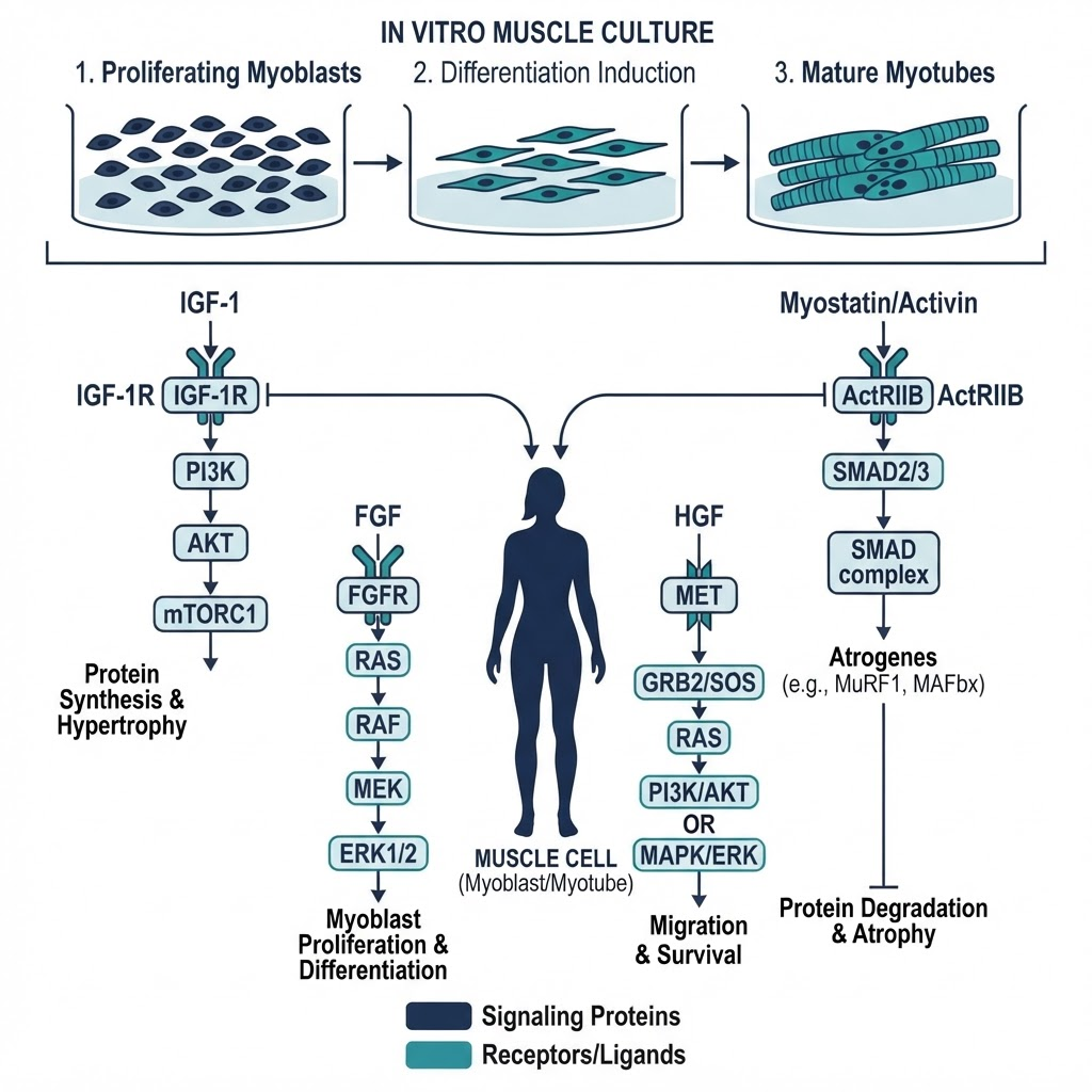
Insulin-like growth factor signaling
IGF-1 and IGF-2 signal primarily through the IGF-1 receptor, a receptor tyrosine kinase. Downstream pathways include PI3K/AKT, which regulates protein synthesis and cell growth; MAPK/ERK, which contributes to proliferation and differentiation; and mTOR, which regulates translation. IGF-1 signaling is therefore central to studies of muscle cell growth and differentiation in vitro, although pathway activation should be confirmed with time-matched phosphoprotein and functional measurements.
Myostatin signaling
Myostatin, also known as GDF-8, is a TGF-beta family member that negatively regulates muscle growth. It signals through activin receptor type IIB and associated ALK4 or ALK5 receptors, leading to SMAD2/3 phosphorylation. Myostatin treatment is commonly used to study inhibition of myoblast proliferation or differentiation. A complete experiment should distinguish direct receptor signaling from later changes in cell number or morphology.
Follistatin and related ligands
Follistatin binds myostatin and related ligands, preventing or modifying their interaction with activin receptors. In cell models, the mechanism can be investigated through ligand-binding assays, receptor phosphorylation, SMAD translocation, and rescue experiments. Results depend on the precise protein or peptide construct, its purity, and the ratio between ligand and binding partner.
Fibroblast growth factor signaling
FGF-2 and related ligands signal through fibroblast growth factor receptors. In muscle cell models, FGF signaling is studied in relation to myoblast proliferation, delayed differentiation, and satellite-cell activation. FGF effects are strongly influenced by serum concentration and extracellular matrix presentation, so both variables should be held constant across treatment groups.
Other signaling peptides
Additional systems include hepatocyte growth factor through MET, platelet-derived growth factor through PDGFR, and VEGF in vascularised or co-culture models. These systems should not be treated as interchangeable: each uses different receptors, kinetics, and assay endpoints.
For related content, see [Cell Signaling research category](/research/cell-signaling).
How do you culture skeletal muscle cells in vitro?
Skeletal muscle cell culture requires defined conditions that preserve phenotype and differentiation capacity. Common models include C2C12 mouse myoblasts, L6 rat myoblasts, immortalised human skeletal muscle cells, and primary donor-derived myoblasts.
| Cell Line | Species | Research Characteristics |
|---|---|---|
| C2C12 | Mouse | Fast-differentiating and well characterised |
| L6 | Rat | Robust and straightforward to maintain |
| HSkM | Human | Human skeletal muscle phenotype |
| Primary myoblasts | Multiple | Donor-dependent biology and variability |
Standard conditions often use DMEM or F-10 supplemented with 10–20% FBS, a defined substrate such as gelatin, collagen, or fibronectin, and incubation at 37°C with 5% CO2. Passage number, seeding density, serum batch, and confluence should be recorded for every experiment.
To induce myotube formation, researchers commonly grow myoblasts to approximately 80–90% confluence and switch to differentiation medium containing DMEM with approximately 2% horse serum. Medium is changed at regular intervals, and myotubes typically become measurable over 3–7 days. Differentiation should be confirmed with myosin heavy chain staining, fusion-index calculation, and, where relevant, myotube diameter measurements.
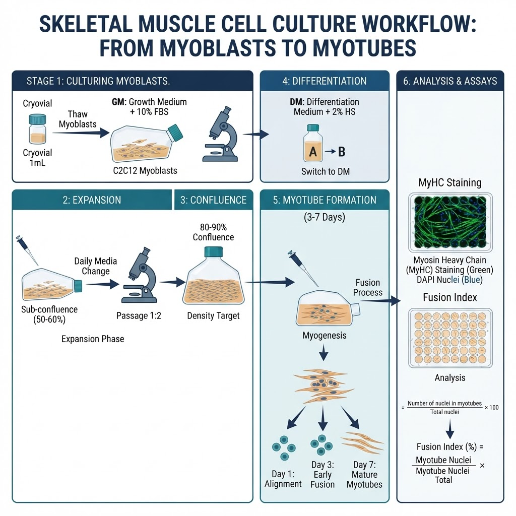
Peptide treatment can be applied during proliferation, early differentiation, late differentiation, or after mature myotubes have formed. The timing must match the research question: an acute phosphorylation experiment should not be interpreted as evidence of a long-term morphological effect.
What receptor systems are studied in muscle cells?
Multiple receptor classes are used to investigate peptide signaling in muscle models.
| Receptor Class | Example Receptor | Ligand | Downstream Pathway |
|---|---|---|---|
| Receptor tyrosine kinase | IGF-1R | IGF-1, IGF-2 | PI3K/AKT, MAPK/ERK |
| Receptor tyrosine kinase | FGFR | FGF-2, FGF-6 | MAPK/ERK, PLC-gamma |
| Receptor tyrosine kinase | MET | HGF | PI3K/AKT, MAPK/ERK |
| Serine/threonine kinase | ActRIIB | Myostatin, activin | SMAD2/3 |
| Serine/threonine kinase | BMPR | BMPs | SMAD1/5/8 |
| G-protein coupled receptor | Beta-2 adrenergic receptor | Beta agonists | cAMP/PKA |
Each receptor system activates distinct downstream pathways that can be measured through time-course experiments, inhibitor studies, immunoblotting, imaging, and functional assays. A receptor antagonist or genetic knockdown can help determine whether an observed response is receptor-dependent.
What analytical methods verify muscle peptide research?
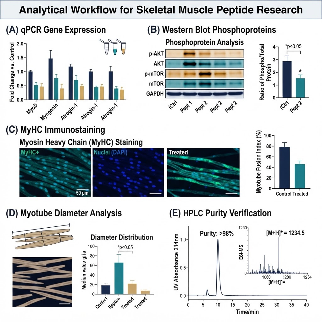
Multiple analytical methods are used to characterise peptide effects on muscle cells. qPCR or RNA sequencing can quantify myogenic markers such as MyoD, myogenin, and MyHC, alongside atrophy markers including MuRF1 and Atrogin-1 and metabolic markers such as PGC-1alpha and GLUT4. Transcript abundance should be interpreted alongside protein and functional data.
Western blotting or ELISA can quantify phosphorylated AKT, ERK, and SMAD proteins, as well as structural and metabolic proteins. For differentiation, MyHC immunostaining and fusion-index analysis provide complementary information. Functional assays may include EdU or BrdU incorporation, resazurin or MTT-based viability measurements, myotube diameter, glucose uptake, and mitochondrial function.
For any peptide used in muscle research, verify identity by mass spectrometry, purity by HPLC, and endotoxin burden where appropriate for cell culture. Batch-specific certificates and storage records should accompany the experiment. See the [HPLC Chromatography Guide](/blog/hplc-chromatography-guide) and [Peptide Purity Testing Standards](/blog/peptide-purity-testing-standards).
What are the key experimental considerations?
Reproducible studies require attention to cell passage and density, serum concentration, treatment duration, and controls. Myoblasts should be maintained within a consistent passage range and seeded at a defined density. Serum contains growth factors that affect differentiation, so growth medium, differentiation medium, and any serum-starvation step should be documented precisely.
Acute phosphorylation events may occur within minutes, gene-expression changes over hours, and differentiation or morphology changes over several days. A useful time-course design can therefore include 5–30 minute signaling points, 6–24 hour transcriptional points, and 3–14 day structural endpoints.
Essential controls include untreated and vehicle controls, a positive control appropriate to the pathway, a scrambled-peptide or inactive-sequence control, and a receptor-antagonist control where available. Researchers should avoid pooling data across peptide batches without demonstrating comparable identity, purity, and activity.
What peptide systems are commonly studied?
IGF-1 and analogues are studied for effects on myoblast proliferation, myotube morphology, and PI3K/AKT/mTOR signaling. Myostatin and follistatin systems are used to examine inhibitory signaling and ligand sequestration. MGF is studied as an IGF-related splice-variant model in satellite-cell and repair research. Growth-hormone secretagogue systems may be investigated indirectly through GH/IGF-1 axis models, but they should not be presented as evidence of a direct muscle-cell mechanism without appropriate receptor and pathway data.
Related materials include [Ipamorelin and CJC-1295: Pharmacological Profile in Preclinical Models](/blog/ipamorelin-cjc-1295-preclinical-profile), the [Repair Signaling Peptides research category](/research/repair-signaling-peptides-overview), and [Peptide Signaling in Dermal Cell Research](/blog/peptide-signaling-dermal-cell-research).
What are common pitfalls in muscle peptide research?
Common errors include passage drift, unverified differentiation, serum-batch variability, uncharacterised peptides, mixing independent batches, inadequate controls, and relying on a single timepoint. Another major pitfall is overinterpreting in-vitro data as evidence of in-vivo muscle function. Cell culture models isolate specific mechanisms; they do not reproduce tissue architecture, immune interactions, vascular supply, or whole-organism pharmacokinetics.
What are the research applications?
Muscle peptide research supports studies of proliferation, differentiation, atrophy signaling, regeneration models, and metabolism. Researchers may use satellite cells and myoblasts to investigate regeneration, or examine glucose uptake, mitochondrial function, and lipid metabolism in differentiated myotubes. The most informative studies connect a molecular endpoint to a validated functional readout and clearly report model limitations.
For related research, see [Mitochondrial Research](/research/mitochondrial-research) and [BPC-157 Cellular Analysis Breakthrough](/blog/bpc-157-cellular-analysis-breakthrough).
Frequently Asked Questions
What are the main peptide signaling systems in skeletal muscle cells?
IGF-1 signaling through IGF-1R, myostatin signaling through ActRIIB, FGF signaling through FGFRs, and HGF signaling through MET are commonly studied systems. Each regulates distinct aspects of muscle-cell biology.
What cell lines are used for skeletal muscle research?
C2C12, L6, immortalised human skeletal muscle cells, and primary human myoblasts are commonly used. Model choice should match the question and be reported with passage and donor details.
How do you induce myotube differentiation in vitro?
Researchers typically grow myoblasts to 80–90% confluence and switch to differentiation medium containing reduced serum. Myotube formation is usually assessed over 3–7 days with MyHC staining and fusion-index analysis.
What analytical methods measure peptide effects on muscle cells?
qPCR, RNA sequencing, Western blotting, ELISA, MyHC staining, myotube measurements, viability assays, and metabolic assays can be combined to measure molecular and functional effects.
What controls are essential for muscle peptide studies?
Untreated, vehicle, pathway-positive, inactive-sequence or scrambled-peptide, and receptor-antagonist controls are useful where technically appropriate.
How should peptides be stored for muscle cell research?
Follow the supplier's documented stability conditions. Lyophilised material is generally stored sealed and protected from moisture, while reconstituted solutions should be aliquoted to minimise repeated freeze–thaw cycles.
Conclusion
Skeletal muscle cells respond to multiple peptide signaling systems that regulate proliferation, differentiation, and metabolism. Myoblast and myotube models provide controlled systems for studying these pathways, but reproducibility depends on consistent culture conditions, analytical verification, appropriate controls, and timepoints matched to the biological question.
All compounds referenced in this article are intended for in-vitro laboratory research only. They are not medicines, supplements, or consumer products, and are not for human or animal use.
References
- Florini JR, Ewton DZ, Coolican SA. (1996). Growth hormone and the insulin-like growth factor system in myogenesis. Endocrine Reviews, 17(5), 481–517.
- McPherron AC, Lawler AM, Lee SJ. (1997). Regulation of skeletal muscle mass in mice by a new TGF-beta superfamily member. Nature, 387(6628), 83–90.
- Glass DJ. (2003). Signalling pathways that mediate skeletal muscle hypertrophy and atrophy. Nature Cell Biology, 5(2), 87–90.
- Charge SB, Rudnicki MA. (2004). Cellular and molecular regulation of muscle regeneration. Physiological Reviews, 84(1), 209–238.
Frequently asked questions
What are the main peptide signaling systems in skeletal muscle cells?
IGF-1R, ActRIIB, FGFR, and MET signaling are commonly studied systems in skeletal muscle cell models.
What cell lines are used for skeletal muscle research?
C2C12, L6, immortalised human skeletal muscle cells, and primary human myoblasts are commonly used.
How do you induce myotube differentiation in vitro?
Myoblasts are commonly grown to 80–90% confluence and transferred to reduced-serum differentiation medium, with formation assessed by MyHC staining and fusion index.
What analytical methods measure peptide effects on muscle cells?
qPCR, RNA sequencing, Western blotting, ELISA, immunostaining, morphology, viability, and metabolic assays can be combined.
What controls are essential for muscle peptide studies?
Untreated, vehicle, pathway-positive, inactive-sequence, and receptor-antagonist controls are useful where technically appropriate.
Frequently Asked Questions
What are the main peptide signaling systems in skeletal muscle cells?
IGF-1R, ActRIIB, FGFR, and MET signaling are commonly studied systems in skeletal muscle cell models.
What cell lines are used for skeletal muscle research?
C2C12, L6, immortalised human skeletal muscle cells, and primary human myoblasts are commonly used.
How do you induce myotube differentiation in vitro?
Myoblasts are commonly grown to 80–90% confluence and transferred to reduced-serum differentiation medium, with formation assessed by MyHC staining and fusion index.
What analytical methods measure peptide effects on muscle cells?
qPCR, RNA sequencing, Western blotting, ELISA, immunostaining, morphology, viability, and metabolic assays can be combined.
What controls are essential for muscle peptide studies?
Untreated, vehicle, pathway-positive, inactive-sequence, and receptor-antagonist controls are useful where technically appropriate.
Dr Sarah Chen
Our research team combines expertise in biochemistry, skincare science, and sports medicine to bring you evidence-based peptide information.
Ready to Explore Peptides?
Discover our selection of research-grade peptides and start your optimisation journey today.
Shop Peptides