Disclaimer: For in-vitro laboratory research only. Not for human or animal use.
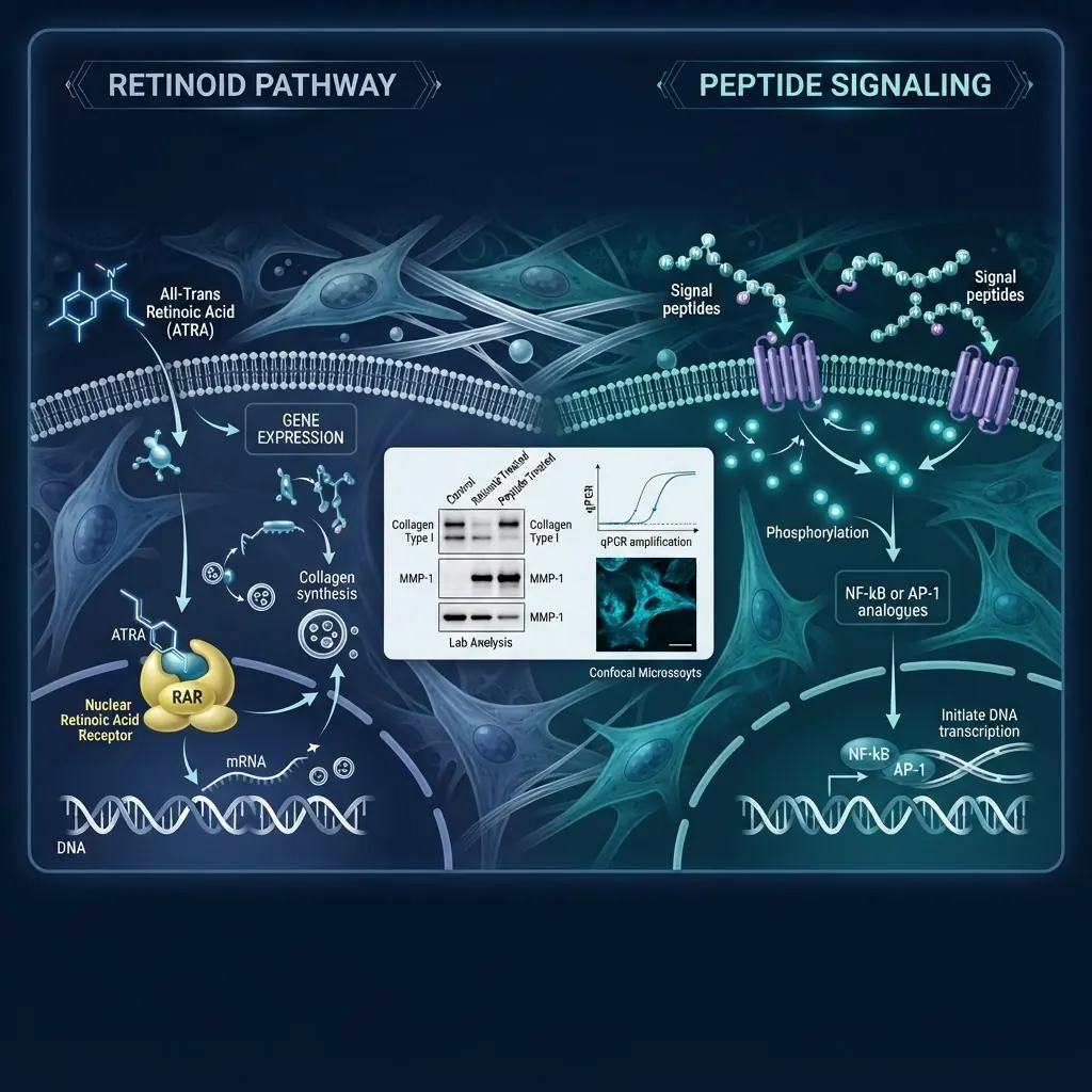
Retinoids and signaling peptides represent two distinct classes of compounds studied in dermal cell research. Retinoids act primarily through nuclear hormone receptors that modulate gene transcription, while signaling peptides typically bind to cell-surface receptors and trigger intracellular phosphorylation cascades. Understanding these differences is essential for designing comparative in-vitro studies.
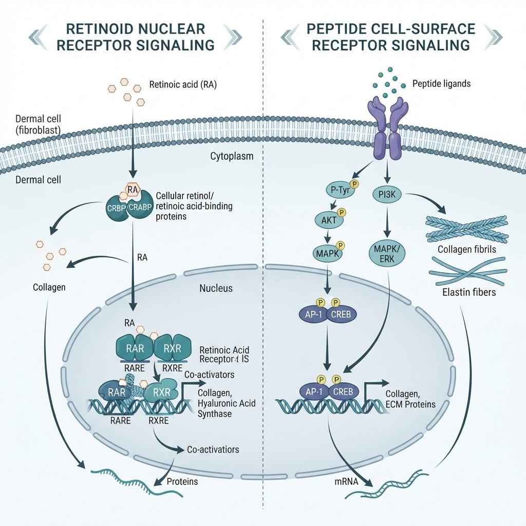
This overview compares molecular mechanisms, receptor systems, downstream pathways, analytical verification, and experimental design. All compounds referenced are intended for in-vitro laboratory research only.
What are retinoids and how do they work in dermal cell research?
Retinoids are compounds structurally related to vitamin A. Common research materials include all-trans retinoic acid (ATRA), retinol, retinaldehyde, and synthetic retinoids. Retinoids act primarily through retinoic acid receptors (RAR) and retinoid X receptors (RXR). RAR-RXR heterodimers bind retinoic acid response elements in DNA and regulate transcription.
Retinoid receptor activation is studied in relation to keratinocyte differentiation, extracellular matrix remodelling, proliferation, apoptosis, and retinoid metabolism. Because transcription and protein synthesis take time, many retinoid readouts are collected over hours to days.
How do signaling peptides work in dermal cell research?
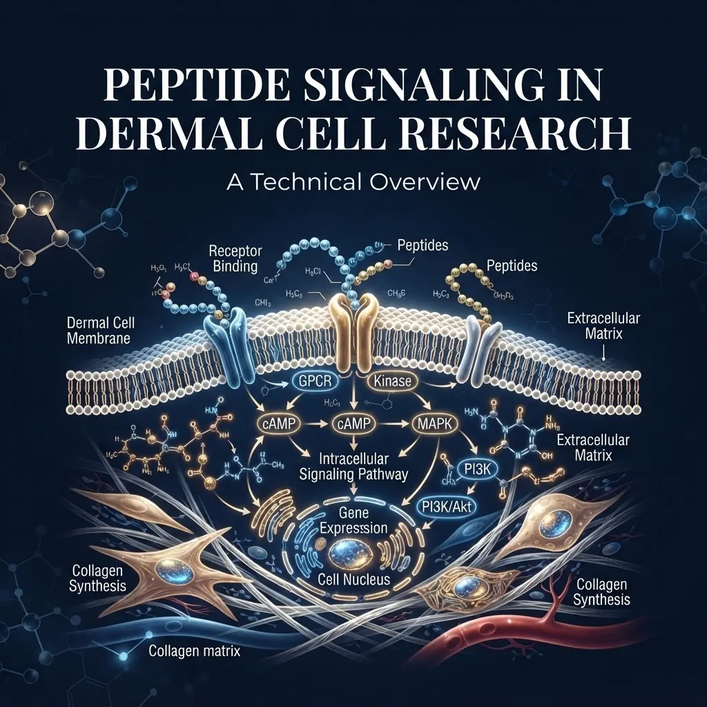
Signaling peptides are short amino acid sequences that bind to cell-surface receptors or interact with extracellular matrix components. Research classes include growth-factor-derived peptides, extracellular-matrix-derived sequences, copper-binding systems such as GHK-Cu, and antimicrobial peptides.
Receptor families investigated in these models include EGFR, FGFR, IGF-1R, and integrins. Their activation can initiate MAPK/ERK, PI3K/AKT, and FAK/Src pathways. Phosphorylation events may be measurable within minutes, followed by immediate-early gene expression and other changes over several hours.
What are the key mechanistic differences?
| Aspect | Retinoids | Signaling peptides |
|---|---|---|
| Receptor location | Nuclear | Cell surface |
| Primary mechanism | Transcription modulation | Phosphorylation cascades |
| Typical early readout | Gene expression | Receptor or kinase phosphorylation |
| Useful timepoints | 24–72 hours | 15 minutes–24 hours |
| Verification priorities | Isomer ratio and purity | Sequence, purity, and content |
These differences mean that a fair comparison cannot rely on one timepoint or one assay. Retinoid studies often require transcriptional readouts, while peptide studies benefit from early phospho-protein measurements and later gene-expression analysis.
How do you design a comparative in-vitro study?
Select a cell model that responds measurably to both compound classes. Dermal fibroblasts and keratinocytes may be suitable, but receptor expression and baseline responsiveness should be confirmed for each model. Use concentration-response curves and prespecified timepoints that capture both rapid peptide signaling and slower retinoid transcriptional effects.
Include untreated, vehicle, positive-pathway, and concentration-response controls. Record cell passage, media composition, plate format, incubation conditions, and compound preparation so that results remain interpretable and reproducible.
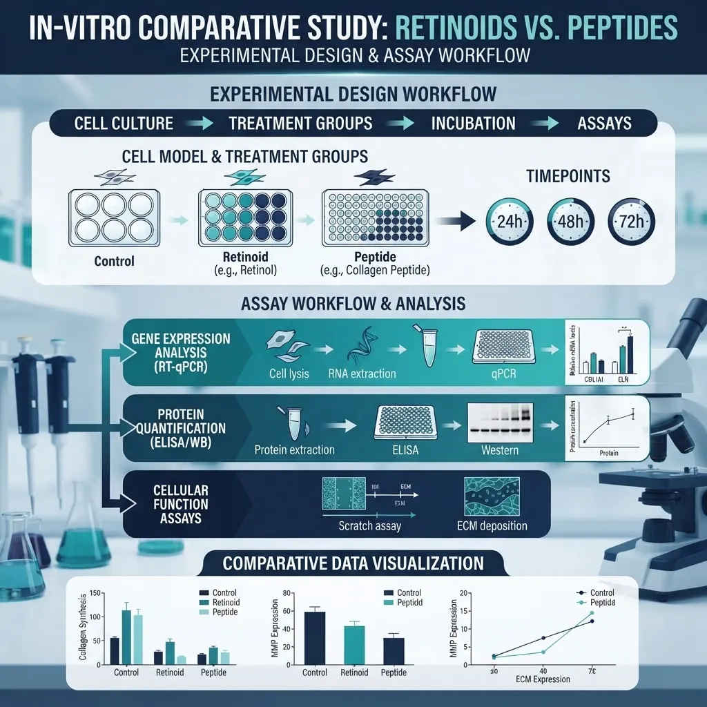
For retinoids, qPCR and protein-expression assays can quantify transcriptional effects. For peptides, phospho-Western, ELISA, and receptor-activation assays can capture early and downstream responses. Proliferation, migration, and extracellular-matrix measurements can provide shared functional endpoints when validated for the model.
What analytical methods verify retinoid and peptide identity?
Retinoids are commonly checked by reverse-phase HPLC with UV detection, mass spectrometry, and isomer-ratio analysis. ATRA can isomerise into 9-cis and 13-cis forms, which may have different receptor selectivity, so light protection and batch-specific verification are important.
Peptides are commonly verified using reverse-phase HPLC, mass spectrometry, amino-acid analysis, and peptide-content assays. Purity alone does not establish identity or biological activity. Researchers should retain the certificate of analysis and verify stability in the intended culture medium.

For practical analytical background, see [HPLC Chromatography Guide](/blog/hplc-chromatography-guide), [Peptide Purity Testing Standards](/blog/peptide-purity-testing-standards), and [Peptide Storage and Stability Best Practices](/blog/peptide-storage-stability-science-backed-best-practices).
What are the stability challenges?
Retinoids are sensitive to light, oxygen, temperature, pH, and solvent conditions. Use appropriate amber handling, validated stock solutions, and stability checks. Peptides can be affected by proteases, adsorption to plastic, oxidation, temperature, and repeated freeze-thaw cycles. Low-binding consumables, aliquoting, and validated storage conditions can reduce variability.
How should comparative data be interpreted?
Do not compare a 30-minute peptide phospho-signal with a 30-minute retinoid transcription assay as if they measured the same biological event. Match the endpoint to the mechanism, compare molar rather than mass concentrations, account for vehicle effects, and use the same cell passage and culture conditions across study arms.
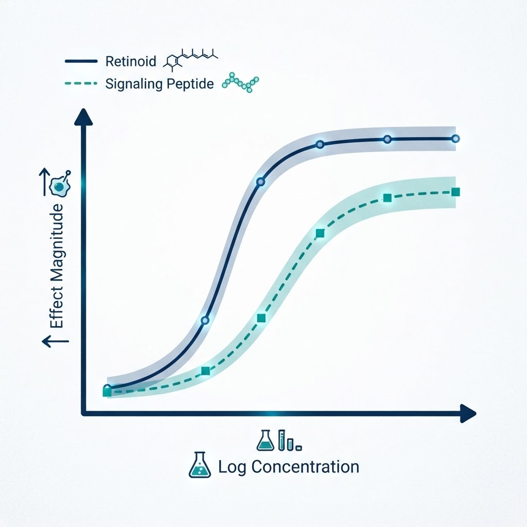
Common pitfalls include mismatched timepoints, photodegradation, peptide adsorption, missing vehicle controls, and single-readout designs. A multiparameter design is more informative than selecting a single assay that favours one compound class.
Related research
- [Peptide Signaling in Dermal Cell Research: A Technical Overview](/blog/peptide-signaling-dermal-cell-research)
- [HPLC Chromatography Guide](/blog/hplc-chromatography-guide)
- [Peptide Purity Testing Standards](/blog/peptide-purity-testing-standards)
- [Peptide Storage and Stability Best Practices](/blog/peptide-storage-stability-science-backed-best-practices)
- [Cell Signaling](/research/cell-signaling)
- [GHK-Cu 50mg Copper Peptide](/product/ghk-cu-50mg-copper-peptide)
Frequently Asked Questions
What is the primary mechanistic difference?
Retinoids primarily regulate transcription through nuclear receptors, while signaling peptides commonly activate cell-surface receptors and phosphorylation cascades.
Can both classes be studied in one experiment?
Yes, if timepoints, controls, concentrations, and readouts are selected for both mechanisms.
Why is ATRA isomerisation important?
Different isomers may have different receptor selectivity, changing the effective pharmacology of the test material.
What controls are essential?
At minimum, include untreated, vehicle, positive-pathway, and concentration-response controls for each compound class.
Conclusion
Retinoids and signaling peptides represent different routes to cellular regulation. Comparative studies should align timepoints, readouts, analytical verification, and controls with each compound's mechanism. All compounds referenced are intended for in-vitro laboratory research only and are not for human or animal use.
References
- Fisher GJ, Voorhees JJ. (1996). Molecular mechanisms of retinoid actions in skin. FASEB Journal, 10(9), 1002–1013.
- Chambon P. (1996). A decade of molecular biology of retinoic acid receptors. FASEB Journal, 10(9), 940–954.
- Bennett NT, Schultz GS. (1993). Growth factors and wound healing. American Journal of Surgery, 165(6), 728–737.
- Werner S, Grose R. (2003). Regulation of wound healing by growth factors and cytokines. Physiological Reviews, 83(3), 835–870.
Frequently asked questions
What is the primary mechanistic difference?
Retinoids primarily regulate transcription through nuclear receptors, while signaling peptides commonly activate cell-surface receptors and phosphorylation cascades.
Can both classes be studied in one experiment?
Yes, if timepoints, controls, concentrations, and readouts are selected for both mechanisms.
What controls are essential?
Include untreated, vehicle, positive-pathway, and concentration-response controls for each compound class.
Frequently Asked Questions
What is the primary mechanistic difference?
Retinoids primarily regulate transcription through nuclear receptors, while signaling peptides commonly activate cell-surface receptors and phosphorylation cascades.
Can both classes be studied in one experiment?
Yes, if timepoints, controls, concentrations, and readouts are selected for both mechanisms.
What controls are essential?
Include untreated, vehicle, positive-pathway, and concentration-response controls for each compound class.
James Whitfield
Our research team combines expertise in biochemistry, skincare science, and sports medicine to bring you evidence-based peptide information.
Ready to Explore Peptides?
Discover our selection of research-grade peptides and start your optimisation journey today.
Shop Peptides