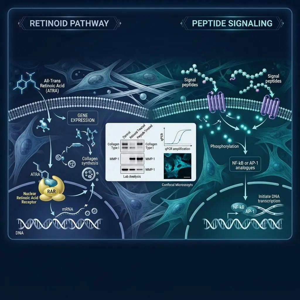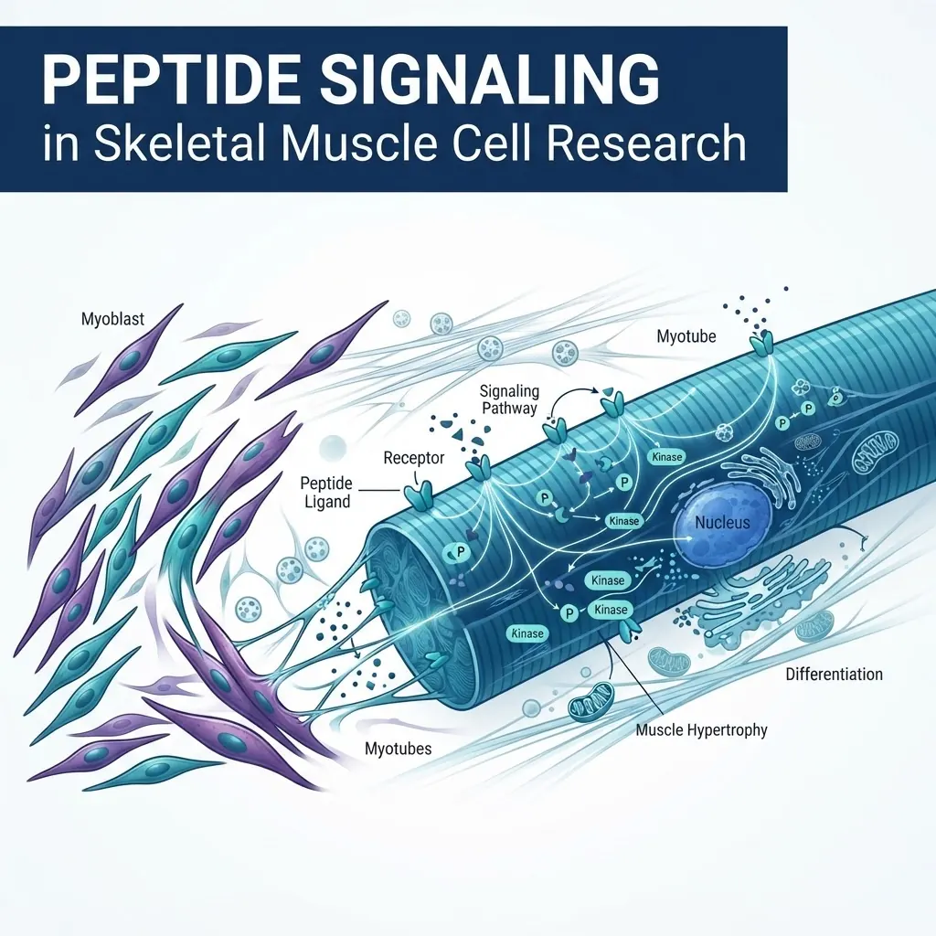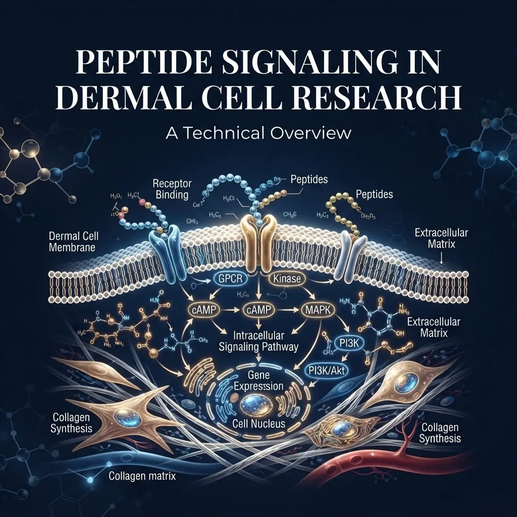
Peptide signaling in dermal cell research refers to the study of short amino acid sequences that bind to cell-surface receptors on fibroblasts and keratinocytes in vitro. These compounds interact with defined receptor families and intracellular cascades. For researchers, mapping these interactions is essential for designing reproducible models and generating interpretable data.

This overview covers signaling peptide classes, receptor families, analytical verification, culture variables, and reproducibility. All compounds referenced are intended for in-vitro laboratory research only.
What are signaling peptides in dermal cell research?
Signaling peptides are short amino acid sequences, commonly ranging from 2 to 50 residues, that bind to receptors or interact with extracellular matrix components. Compared with full-length proteins, they offer defined sequence, known molecular weight, reproducible synthesis, and controlled molar application in cell models.
Growth factor-derived peptides
Fragments derived from epidermal growth factor, fibroblast growth factor, or insulin-like growth factor sequences may retain selected receptor-binding characteristics while being easier to characterise than full recombinant proteins. Researchers should confirm the sequence, folding requirements, purity, and functional activity for each construct rather than assuming that a fragment reproduces the parent protein.
Extracellular matrix-derived peptides
Collagen, elastin, and fibronectin fragments are studied for interactions with integrins and other adhesion-associated receptors. Common research examples include collagen-derived tripeptides, elastin-derived sequences, and RGD motifs. These models can be used to investigate adhesion, migration, focal adhesion kinase activity, and matrix remodelling.
Copper-binding peptides
GHK-Cu, or glycyl-L-histidyl-L-lysine copper complex, is studied as a copper-binding system rather than as a conventional receptor tyrosine kinase ligand. Its experimental interpretation depends on confirming complex formation, copper stoichiometry, and the stability of the prepared material.
Antimicrobial peptides
Cationic peptides such as LL-37 interact with membranes and are used in models of innate immune signaling. Their effects may depend on concentration, ionic strength, serum composition, and membrane integrity, so controls are particularly important.
How do peptide-receptor interactions work in dermal cell models?
Peptide-receptor binding follows standard ligand-receptor kinetics. Frequently studied systems include EGFR with EGF-derived sequences, FGFR with FGF fragments, IGF-1R with IGF analogues, and integrins with extracellular-matrix peptides.

| Receptor family | Example ligand | Downstream pathway | Typical readout |
|---|---|---|---|
| EGFR | EGF-derived peptides | MAPK/ERK | Phospho-ERK Western blot |
| FGFR | FGF fragments | PI3K/AKT | Phospho-AKT ELISA |
| IGF-1R | IGF analogues | MAPK and PI3K | Receptor autophosphorylation |
| Integrins | ECM-derived peptides | FAK/Src | Adhesion assays |
| Membrane receptors | LL-37 | NF-κB | Cytokine assays |
Binding affinity can be examined using surface plasmon resonance, isothermal titration calorimetry, radioligand binding, or cell-based reporter assays. Direct methods provide kinetic or thermodynamic parameters; cell assays provide functional confirmation but may combine receptor effects with uptake, degradation, and secondary signaling.
Receptor specificity should be tested rather than inferred. Useful controls include receptor-specific inhibitors, siRNA knockdown, comparison with full-length ligands, scrambled sequences, and dose-response curves across receptor subtypes. Cross-reactivity should be reported because it changes the interpretation of downstream measurements.
What analytical methods verify peptide identity and purity?
Before a signaling study, verify identity, purity, concentration, and—where relevant—endotoxin content. Batch-specific documentation is more useful than a generic specification sheet.
High-performance liquid chromatography
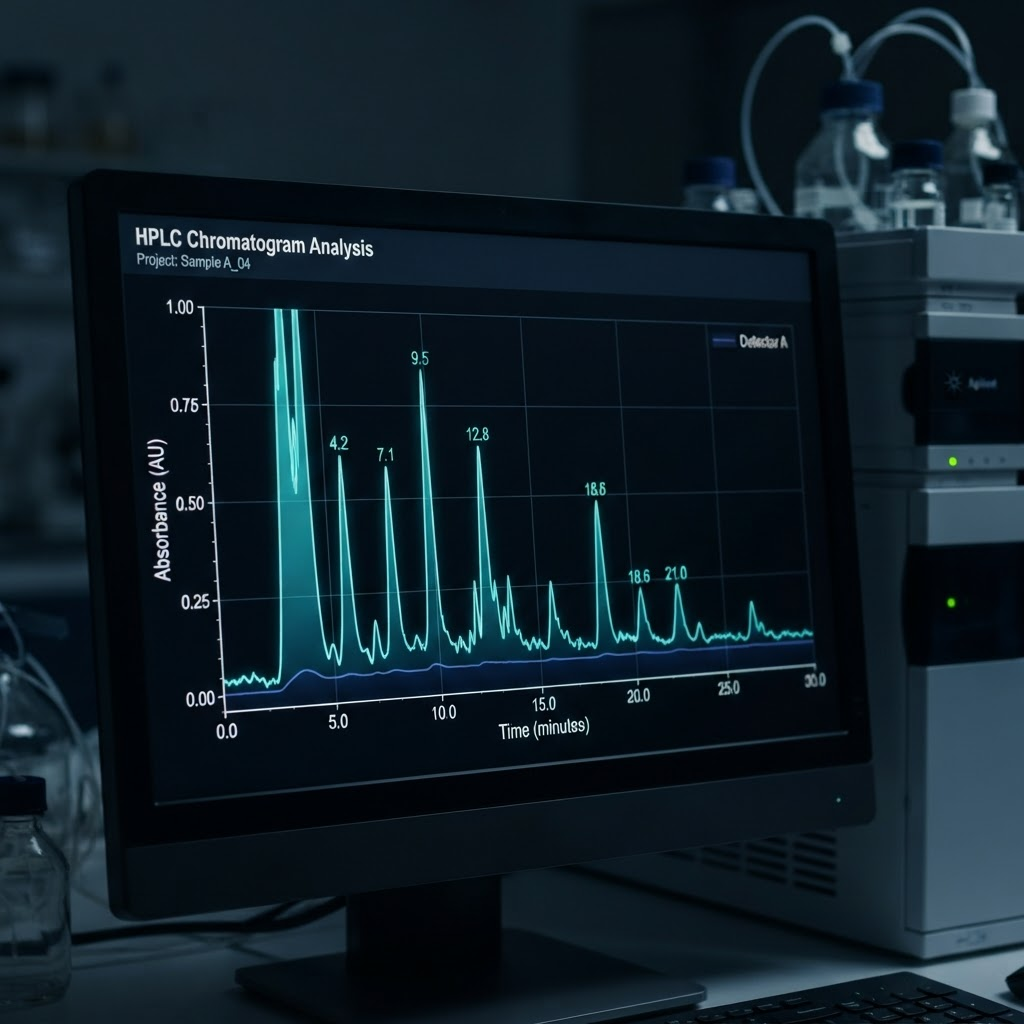
Reverse-phase HPLC separates peptides by hydrophobicity and reports purity as the target peak area relative to total detected area. A C18 column, water/acetonitrile gradient, acidic modifier, and UV detection near 214 nm are common starting conditions. The method should be validated for the sequence and matrix being tested.
Mass spectrometry
Mass spectrometry confirms molecular weight and, with tandem analysis, provides sequence information. Electrospray ionisation is useful for many dissolved peptides, while MALDI-TOF offers rapid mass confirmation. Observed mass should be compared with the theoretical mass while accounting for counter-ions and modifications.
Content, amino acid, and endotoxin testing
A content assay distinguishes peptide mass from total lyophilised powder, which may include water, acetate, or TFA. Amino acid analysis provides compositional confirmation. For cell culture, endotoxin testing is an important control because contaminating endotoxin can activate inflammatory pathways independently of the test peptide.
For copper complexes, add copper quantification, UV-visible stability measurements, and stoichiometric analysis. Free peptide and peptide-copper complex should not be treated as interchangeable materials.
What variables affect dermal fibroblast culture studies?
Passage number, serum batch, media composition, plating density, and incubation time can all change signaling responses. Many laboratories use a defined passage window and record it for every experiment. Serum reduction before stimulation can lower baseline pathway activity, but the starvation conditions must be empirically established for each cell model.
| Pathway or endpoint | Typical observation window |
|---|---|
| MAPK/ERK phosphorylation | 5–30 minutes |
| PI3K/AKT phosphorylation | 15–60 minutes |
| Transcriptional response | 6–24 hours |
| Protein abundance | 24–72 hours |
A time course is preferable to selecting one timepoint from a different model. Common readouts include Western blot, ELISA, reporter assays, immunofluorescence, and qPCR. Every experiment should include untreated, vehicle, positive, and scrambled-sequence controls where appropriate.
Independent concentration verification is also valuable. Depending on sequence, researchers may use UV absorbance, colorimetric protein assays, or amino acid analysis. Stated mass concentration can overestimate active peptide when powder content or counter-ion composition is not considered.
How do copper-binding peptides differ from growth factor peptides?
Copper-binding systems are studied through metal coordination, gene-expression changes, and extracellular-matrix-associated endpoints, while growth factor fragments are usually evaluated through direct receptor-mediated phosphorylation. GHK-Cu can be examined in relation to lysyl oxidase, superoxide dismutase, cytochrome c oxidase, matrix metalloproteinases, and collagen-associated transcripts.
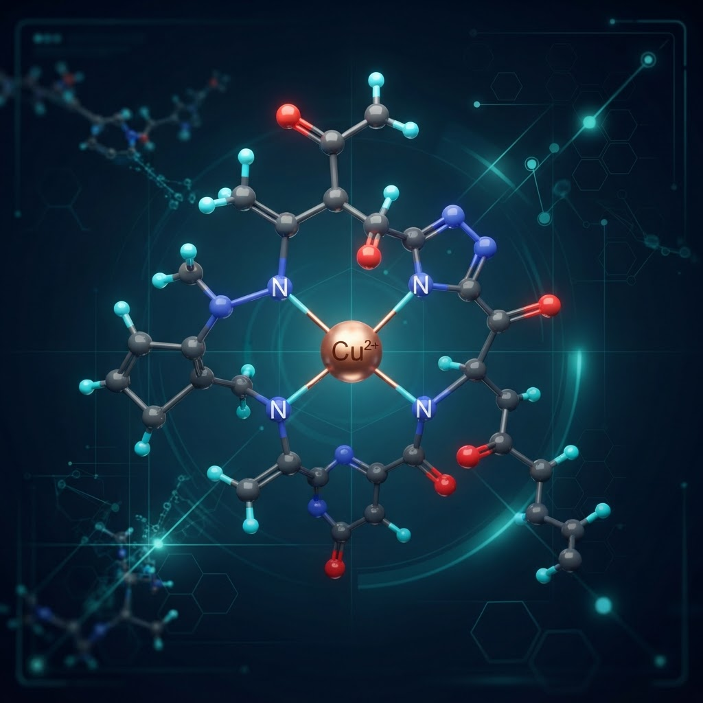
These experiments require careful controls for free copper, unconjugated peptide, and vehicle. Researchers should also specify whether the material was prepared as a defined complex and whether the complex remained stable during incubation. Effects measured over hours or days should not be compared directly with rapid phosphorylation events without considering the different biological time scales.
What stability issues affect peptide signaling studies?
Proteases, temperature, pH, adsorption to plastic, oxidation, and light exposure can reduce the intact concentration in culture. Pre-incubation stability testing can be performed by incubating the material in the intended medium, sampling across a time course, and analysing intact peptide by HPLC. This establishes whether the nominal concentration remains a useful estimate.
Lyophilised material should be kept sealed under suitable low-temperature conditions. Reconstituted solutions should be aliquoted to limit freeze-thaw events, and sequence-specific oxidation risks should be recorded. Low-binding plates and compatible buffers may reduce adsorption, but mitigation should be demonstrated rather than assumed.
How should peptide research data be reported?
A reproducible report should include peptide sequence, molecular weight, modifications, HPLC purity, mass-spectrometry confirmation, lot number, supplier, reconstitution solvent, concentration verification method, cell source, passage number, serum conditions, treatment time, controls, sample size, and statistical method. Include representative analytical traces when possible.
Frequently Asked Questions
What purity level is required for dermal cell signaling studies?
Many in-vitro signaling studies specify at least 95% HPLC purity, with higher purity preferred for quantitative binding work. The suitable threshold depends on the assay and should be justified.
How do I verify peptide identity?
Request batch-specific HPLC and mass-spectrometry data, compare measured and theoretical mass, and use amino acid analysis where compositional confirmation is needed.
What concentration range is typical?
Published in-vitro models vary widely, often spanning nanomolar to micromolar concentrations. Establish a dose-response curve for the specific receptor and cell model rather than transferring a concentration without validation.
Are copper-binding peptides used differently from growth factor peptides?
Yes. Copper complexes require additional assessment of metal content, complex stability, and free-copper controls, while growth-factor-derived sequences are often examined through receptor phosphorylation.
What are the main sources of variability?
Passage number, serum batch, media, plating density, peptide stability, endotoxin, and inaccurate concentration estimates are common sources of variation.
How long should cultures be exposed?
The appropriate window depends on the endpoint. Phosphorylation may change within minutes, transcription over hours, and protein abundance over one to three days. A time course is recommended.
What controls are essential?
At minimum, include untreated and vehicle controls plus positive and sequence-matched negative controls where appropriate. Receptor-specific inhibition or knockdown can strengthen mechanism claims.
How should peptides be stored?
Store sealed lyophilised material under suitable low-temperature conditions and aliquot reconstituted solutions to reduce freeze-thaw exposure. Validate sequence-specific stability in the intended medium.
Conclusion
Peptide signaling in dermal cell research is best approached as a measurement problem: define the ligand, verify the material, control the cell model, select a time course, and report the analytical evidence. These practices help distinguish receptor-mediated effects from degradation, contamination, matrix effects, and assay artefacts. All compounds referenced here are intended for in-vitro laboratory research only and are not for human or animal use.
References
- Pickart L, Margolina A. (2018). Regenerative and protective actions of the GHK-Cu peptide. Biomedical Reports, 8(5), 405–411.
- Bennett NT, Schultz GS. (1993). Growth factors and wound healing: biochemical properties of growth factors and their receptors. American Journal of Surgery, 165(6), 728–737.
- Werner S, Grose R. (2003). Regulation of wound healing by growth factors and cytokines. Physiological Reviews, 83(3), 835–870.
- Lintner K, Peschard O. (2000). Biologically active peptides. International Journal of Cosmetic Science, 22(3), 207–218.
All compounds referenced are intended for in-vitro laboratory research only.
Frequently asked questions
What purity level is required for dermal cell signaling studies?
Many in-vitro signaling studies specify at least 95% HPLC purity, with higher purity preferred for quantitative binding work.
How do I verify peptide identity?
Request batch-specific HPLC and mass-spectrometry data, compare measured and theoretical mass, and use amino acid analysis where needed.
What concentration range is typical?
Published in-vitro models vary widely, often spanning nanomolar to micromolar concentrations. Establish a dose-response curve for the specific model.
Are copper-binding peptides used differently from growth factor peptides?
Yes. Copper complexes require assessment of metal content, complex stability, and free-copper controls.
What are the main sources of variability?
Passage number, serum batch, media, plating density, peptide stability, endotoxin, and concentration estimates.
How long should cultures be exposed?
The appropriate window depends on the endpoint; phosphorylation, transcription, and protein abundance occur on different time scales.
What controls are essential?
Include untreated, vehicle, positive, and suitable sequence-matched negative controls.
How should peptides be stored?
Store sealed lyophilised material under suitable low-temperature conditions and aliquot reconstituted solutions.
Frequently Asked Questions
What purity level is required for dermal cell signaling studies?
Many in-vitro signaling studies specify at least 95% HPLC purity, with higher purity preferred for quantitative binding work.
How do I verify peptide identity?
Request batch-specific HPLC and mass-spectrometry data, compare measured and theoretical mass, and use amino acid analysis where needed.
What concentration range is typical?
Published in-vitro models vary widely, often spanning nanomolar to micromolar concentrations. Establish a dose-response curve for the specific model.
Are copper-binding peptides used differently from growth factor peptides?
Yes. Copper complexes require assessment of metal content, complex stability, and free-copper controls.
What are the main sources of variability?
Passage number, serum batch, media, plating density, peptide stability, endotoxin, and concentration estimates.
How long should cultures be exposed?
The appropriate window depends on the endpoint; phosphorylation, transcription, and protein abundance occur on different time scales.
What controls are essential?
Include untreated, vehicle, positive, and suitable sequence-matched negative controls.
How should peptides be stored?
Store sealed lyophilised material under suitable low-temperature conditions and aliquot reconstituted solutions.
Dr. Sarah Chen
Our research team combines expertise in biochemistry, skincare science, and sports medicine to bring you evidence-based peptide information.
Ready to Explore Peptides?
Discover our selection of research-grade peptides and start your optimisation journey today.
Shop Peptides