Disclaimer: This article is for qualified laboratory researchers and institutions. It discusses research compounds, specifications, analytical methods, and in-vitro study context only. It does not provide human or animal use instructions.
Age-related changes in connective tissue are characterised by altered extracellular matrix (ECM) composition, lower collagen synthesis, increased matrix metalloproteinase activity, and accumulation of advanced glycation end products. Collagen peptide fragments are used in laboratory research to study these processes in controlled in-vitro models. This overview covers age-related ECM biology, model design, fragment signaling, and analytical verification.
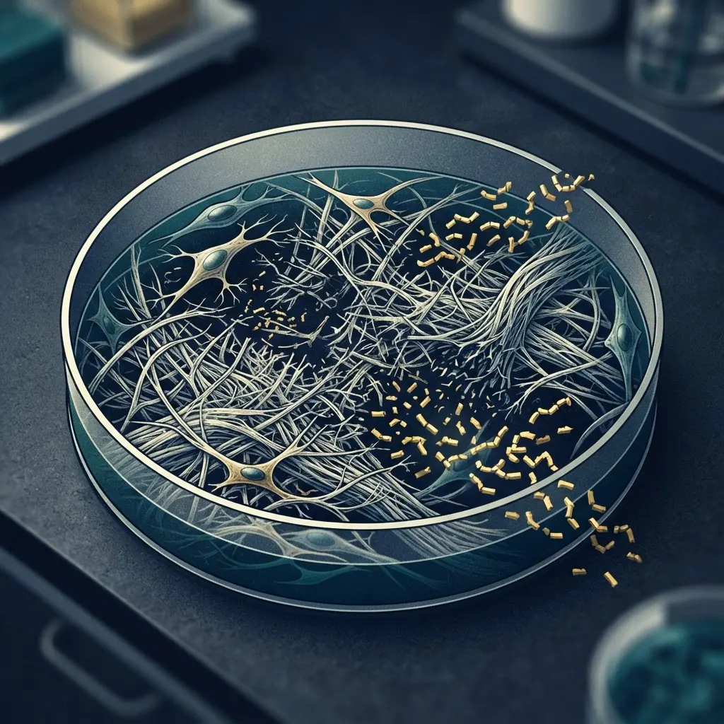
All materials referenced in this article are intended for in-vitro laboratory research only.
What are the age-related changes in connective tissue?
Ageing alters connective tissue through several interacting mechanisms. Fibroblast collagen synthesis can decline with cellular age, which may appear in vitro as reduced COL1A1 and COL3A1 expression, lower procollagen secretion, and decreased collagen deposition. Results depend on donor source, passage number, substrate, oxygen tension, and culture medium, so those variables should be documented.
Matrix metalloproteinases degrade collagen and other ECM components. Age-related models commonly examine MMP-1, MMP-2, and MMP-9 using zymography, activity assays, immunoassays, or gene-expression analysis. Transcript abundance does not necessarily indicate proportional enzyme activity, making orthogonal measurement important.
Advanced glycation end products form through non-enzymatic modification of collagen lysine and arginine residues. These modifications can increase matrix stiffness and change cell-matrix interactions. Researchers use fluorescence spectroscopy, immunoassays for CML or pentosidine, and mass spectrometry to characterise glycated material. Enzymatic lysyl oxidase crosslinking and non-enzymatic glycation produce different matrix signatures, so studies should report which mechanism is being modelled.
For related structural context, see [Collagen Peptide Fragments in Connective Tissue Research](/blog/collagen-peptide-fragments-connective-tissue).
How do collagen peptide fragments interact with aged cells?
Collagen fragments may interact with cells through adhesion receptors and through changes to the surrounding matrix. Their observed effect depends on sequence, concentration basis, exposure time, cell state, and the analytical endpoint selected by the study.
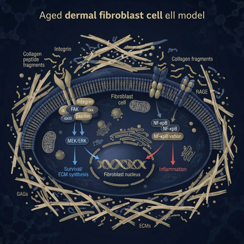
Fragments containing the GFOGER motif can bind collagen-recognising integrins such as alpha-1-beta-1 and alpha-2-beta-1. Cellular age may alter integrin abundance, focal adhesion organisation, and downstream kinase activity. Adhesion assays, immunofluorescence, and phosphoprotein measurements can help distinguish receptor binding from secondary matrix effects.
A collagen fragment can also change the local presentation of matrix ligands or influence how cells organise their substrate. Receptor tyrosine kinase and focal adhesion signaling may therefore be studied alongside matrix composition. Advanced glycation products can engage RAGE and alter inflammatory signaling. Senescent cells may produce a senescence-associated secretory phenotype containing cytokines, MMPs, and growth factors.
How do you design in-vitro models of age-related connective tissue?
Replicative senescence models use cells passaged until they develop a senescence phenotype. Researchers commonly monitor reduced proliferation, increased SA-beta-galactosidase activity, altered p16 and p21 expression, and selected secreted markers. Passage number and culture history must be reported because a passage threshold is not equivalent across cell sources.
Stress-induced models use sublethal oxidative, ultraviolet, or chemical stress to produce a faster phenotype. This is useful for screening, but may not reproduce all features of replicative ageing. The stressor, exposure window, washout conditions, and recovery period should be reported with the marker panel.
Glycation models expose collagen or reconstituted ECM to reducing sugars or reactive carbonyl compounds before cell seeding. This models matrix modification rather than cellular age. A factorial design can compare young cells on young matrix, young cells on aged matrix, aged cells on young matrix, and aged cells on aged matrix. Co-culture or conditioned-medium systems can examine paracrine effects while separating direct contact from secreted factors.
What collagen peptide fragments are used in ageing research?
Selection depends on the research question and the receptor or matrix feature being examined.
| Fragment | Source context | Research application |
|---|---|---|
| Gly-Pro-Hyp | Type I collagen | Fibroblast adhesion and proliferation |
| Pro-Hyp | Type I collagen | Fibroblast migration |
| Hyp-Gly | Type I collagen | Collagen-associated signaling studies |
| GFOGER | Type I collagen | Integrin-mediated adhesion |
| DGEA | Type I collagen | Alpha-2-beta-1 integrin binding |
| CB fragments | Type II collagen | Chondrocyte and cartilage models |
Sequence identity alone is not sufficient to define a research preparation. Purity, counterions, residual process materials, concentration basis, and batch-specific analytical data should also be recorded. For broader context, see [Collagen Peptide Fragments in Connective Tissue Research](/blog/collagen-peptide-fragments-connective-tissue).
What analytical methods characterise age-related changes?
A defensible package combines methods with different sources of selectivity. qPCR or RNA sequencing can quantify collagen, MMP, TIMP, and senescence-associated transcripts. Western blot, ELISA, or targeted proteomics can assess protein abundance. Transcript changes should not be presented as direct evidence of matrix deposition or enzyme activity without the corresponding protein or functional assay.
Hydroxyproline assays estimate total collagen-associated content, while crosslink analysis can examine pyridinoline or deoxypyridinoline. Fluorescence measurements can support AGE analysis, but fluorescence is not a substitute for specific identification when multiple fluorescent species may be present.
SA-beta-galactosidase staining, EdU or BrdU incorporation, scratch or transwell migration, apoptosis assays, and secreted cytokine measurements can be combined to describe cell state. Multiple markers are preferable to a single senescence readout.

For chromatography and purity guidance, see [HPLC Chromatography Guide](/blog/hplc-chromatography-guide) and [Peptide Purity Testing Standards](/blog/peptide-purity-testing-standards).
What are the key experimental considerations?
Primary cells from multiple donors can provide useful biological variation, while replicative models offer tighter control over passage history. Either approach requires a documented cell source, passage number, medium, serum concentration, oxygen condition, substrate, and timepoint.
Essential controls include young and aged cell references, vehicle controls, and positive and negative controls for the selected senescence model. Collagen fragment identity and purity should be verified before exposure studies, with concentration reported on a defined basis. Researchers should record adsorption, precipitation, pH drift, and stability in the actual assay medium.
Donor variability is especially important. Results from one donor or one cell line should be described as model-specific. Matching passage numbers across experimental arms and using independent biological replicates helps separate age-related effects from handling differences.
What are the applications in age-related research?
Collagen peptide fragments are used in fibroblast adhesion, proliferation, migration, matrix-remodelling, senescence, and tissue-engineering models. Scratch assays can examine migration in aged-cell cultures, while three-dimensional scaffolds can test how matrix composition changes cell organisation. These are research endpoints, not evidence of a human-use outcome.
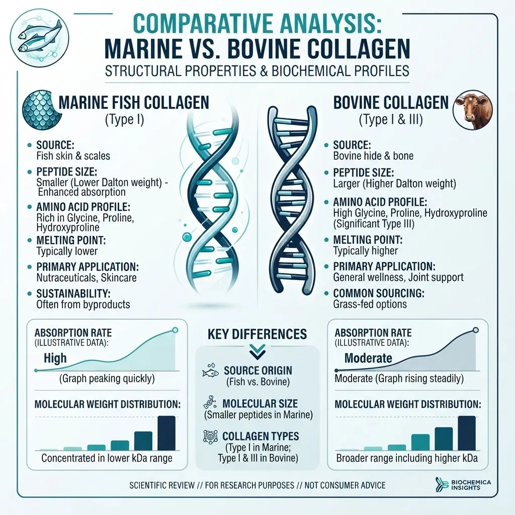
Comparative source studies can examine marine and bovine collagen materials in matched models. See [Marine vs. Bovine Collagen: Source Comparison for Laboratory Studies](/blog/marine-vs-bovine-collagen-lab-comparison) and [Marine Collagen Peptide Fractions: Analytical Comparison](/blog/marine-collagen-peptide-fractions-analysis).
What are common pitfalls in age-related collagen research?
Common errors include undocumented donor age, mixed passage comparisons, single-donor conclusions, inconsistent oxygen or serum conditions, inadequate senescence confirmation, uncharacterised fragments, pooled batches without equivalence testing, and overinterpretation of in-vitro data. A clear study record should connect each result to its cell source, matrix condition, fragment identity, analytical method, and control group.
What are emerging research directions?
Current directions include fraction-specific studies, glycation-modulated models, multi-omics analysis, and matched comparisons of cellular and matrix ageing. Combining transcriptomics, proteomics, metabolomics, and mechanical testing may provide a more complete picture of how aged cells respond to defined collagen fragments.
Related research and products
Related reading includes [Hydrolysed Collagen Peptides: Molecular Structure and Research Applications](/blog/hydrolysed-collagen-peptides-structure), [HPLC Chromatography Guide](/blog/hplc-chromatography-guide), and [Peptide Purity Testing Standards](/blog/peptide-purity-testing-standards). Researchers can also review [Repair Signaling Peptides](/research/repair-signaling-peptides-overview), [Longevity Research](/research/longevity-research), and [Cell Signaling](/research/cell-signaling).
Product pages relevant to laboratory planning include [BPC-157 5mg](/product/bpc-157-5mg), [TB-500 5mg](/product/thymosin-beta-4-5mg-tb500), [GHK-Cu 50mg Copper Peptide](/product/ghk-cu-50mg-copper-peptide), [Bacteriostatic Mixing Water](/product/10ml-bacteriostatic-mixing-water), and [Acetic Acid 0.6% Peptide Solvent](/product/10ml-acetic-acid-0-6-percent-peptide-solvent).
Frequently asked questions
What in-vitro models are used for age-related collagen research?
Replicative senescence, stress-induced senescence, glycation, matrix-ageing, and co-culture models each capture different aspects of connective tissue ageing.
How do you confirm senescence in an in-vitro model?
Use multiple markers, such as SA-beta-galactosidase activity, p16 and p21 expression, reduced proliferation, and selected SASP cytokines.
What collagen fragments are studied in age-related research?
Gly-Pro-Hyp, Pro-Hyp, Hyp-Gly, GFOGER, and DGEA are examples used in studies of collagen-associated signaling and cell-matrix interactions.
How do aged fibroblasts respond differently to collagen fragments?
They may show altered integrin expression, signaling, proliferation, MMP secretion, and matrix deposition. Responses remain dependent on the cell model and assay conditions.
What analytical methods verify age-related changes?
qPCR or RNA sequencing, immunoassays, hydroxyproline measurement, AGE fluorescence, mass spectrometry, and cellular senescence assays can be combined.
How do you control donor variability?
Use multiple donors where possible, document donor age and passage, match culture conditions, and report cell source and replicate structure.
What controls are essential?
Young and aged cell references, vehicle controls, positive and negative model controls, and analytical verification of the collagen fragment preparation are essential.
What are common pitfalls?
Unverified cell age, mixed passage comparisons, insufficient senescence markers, uncharacterised fragments, inconsistent culture conditions, and extrapolation beyond the in-vitro model.
Conclusion
Age-related connective tissue research involves changes in collagen synthesis, matrix degradation, crosslinking, glycation, and cell-state signaling. Collagen peptide fragments provide defined inputs for in-vitro studies, but conclusions depend on model design, donor and passage documentation, orthogonal analytical verification, and appropriate controls.
All materials referenced in this article are intended for in-vitro laboratory research only. They are not medicines, supplements, or consumer products, and are not for human or animal use.
References
- Fisher GJ, Varani J, Voorhees JJ. (2008). Looking older: fibroblast collapse and therapeutic implications. Archives of Dermatology, 144(5), 666–672.
- Varani J, Dame MK, Rittie L, et al. (2006). Decreased collagen production in chronologically aged skin. American Journal of Pathology, 168(6), 1861–1868.
- Verzijl N, DeGroot J, Thorpe SR, et al. (2000). Effect of collagen turnover on the accumulation of advanced glycation end products. Journal of Biological Chemistry, 275(50), 39027–39031.
- Shoulders MD, Raines RT. (2009). Collagen structure and stability. Annual Review of Biochemistry, 78, 929–958.
Frequently asked questions
What in-vitro models are used for age-related collagen research?
Replicative senescence, stress-induced senescence, glycation, matrix-ageing, and co-culture models each capture different aspects of connective tissue ageing.
How do you confirm senescence in an in-vitro model?
Use multiple markers, such as SA-beta-galactosidase activity, p16 and p21 expression, reduced proliferation, and selected SASP cytokines.
What collagen fragments are studied in age-related research?
Gly-Pro-Hyp, Pro-Hyp, Hyp-Gly, GFOGER, and DGEA are examples used in studies of collagen-associated signaling and cell-matrix interactions.
How do aged fibroblasts respond differently to collagen fragments?
They may show altered integrin expression, signaling, proliferation, MMP secretion, and matrix deposition. Responses remain dependent on the cell model and assay conditions.
What analytical methods verify age-related changes?
qPCR or RNA sequencing, immunoassays, hydroxyproline measurement, AGE fluorescence, mass spectrometry, and cellular senescence assays can be combined.
How do you control donor variability?
Use multiple donors where possible, document donor age and passage, match culture conditions, and report cell source and replicate structure.
What controls are essential?
Young and aged cell references, vehicle controls, positive and negative model controls, and analytical verification of the collagen fragment preparation are essential.
What are common pitfalls?
Unverified cell age, mixed passage comparisons, insufficient senescence markers, uncharacterised fragments, inconsistent culture conditions, and extrapolation beyond the in-vitro model.
Frequently Asked Questions
What in-vitro models are used for age-related collagen research?
Replicative senescence, stress-induced senescence, glycation, matrix-ageing, and co-culture models each capture different aspects of connective tissue ageing.
How do you confirm senescence in an in-vitro model?
Use multiple markers, such as SA-beta-galactosidase activity, p16 and p21 expression, reduced proliferation, and selected SASP cytokines.
What collagen fragments are studied in age-related research?
Gly-Pro-Hyp, Pro-Hyp, Hyp-Gly, GFOGER, and DGEA are examples used in studies of collagen-associated signaling and cell-matrix interactions.
How do aged fibroblasts respond differently to collagen fragments?
They may show altered integrin expression, signaling, proliferation, MMP secretion, and matrix deposition. Responses remain dependent on the cell model and assay conditions.
What analytical methods verify age-related changes?
qPCR or RNA sequencing, immunoassays, hydroxyproline measurement, AGE fluorescence, mass spectrometry, and cellular senescence assays can be combined.
How do you control donor variability?
Use multiple donors where possible, document donor age and passage, match culture conditions, and report cell source and replicate structure.
What controls are essential?
Young and aged cell references, vehicle controls, positive and negative model controls, and analytical verification of the collagen fragment preparation are essential.
What are common pitfalls?
Unverified cell age, mixed passage comparisons, insufficient senescence markers, uncharacterised fragments, inconsistent culture conditions, and extrapolation beyond the in-vitro model.
James Whitfield
Our research team combines expertise in biochemistry, skincare science, and sports medicine to bring you evidence-based peptide information.
Ready to Explore Peptides?
Discover our selection of research-grade peptides and start your optimisation journey today.
Shop Peptides