Collagen peptide fragments are short amino acid sequences derived from the hydrolysis or enzymatic cleavage of native collagen proteins. In connective tissue research, these fragments are studied for their role in extracellular matrix signalling, cell adhesion, and matrix remodelling in in-vitro models. Unlike intact collagen, peptide fragments are soluble, characterisable, and suitable for controlled laboratory experiments.
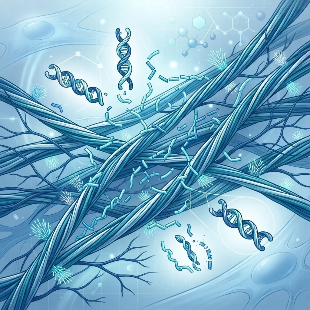
This overview covers the molecular structure of collagen-derived peptides, their interaction with cell-surface receptors, in-vitro research models, analytical verification methods, and experimental design considerations. All compounds referenced are intended for in-vitro laboratory research only.
What are collagen peptide fragments?
Collagen peptide fragments are short chains of amino acids produced by hydrolysis of native collagen. They typically range from two to fifty residues, with molecular weights between approximately 200 Da and 5 kDa. Their properties depend on sequence, source protein, hydrolysis conditions, and purification method.
Structural characteristics
Native collagen is a triple-helical protein composed of repeating Gly-X-Y sequences, where X is often proline and Y is often hydroxyproline. Hydrolysis cleaves the triple helix into smaller fragments with distinct analytical and biological properties.
| Fragment type | Typical length | Research characteristic |
|---|---|---|
| Dipeptides | 2 residues | Small sequence-defined fragments |
| Tripeptides | 3 residues | Frequently used in transport and binding studies |
| Oligopeptides | 4–10 residues | Candidate receptor-active sequences |
| Polypeptides | 10–50 residues | May retain partial structural features |
The biological activity of a collagen fragment depends on its sequence and presentation. Gly-Pro-Hyp and Pro-Hyp are among the most studied small fragments, while GFOGER is widely used in integrin-binding research.
Source materials
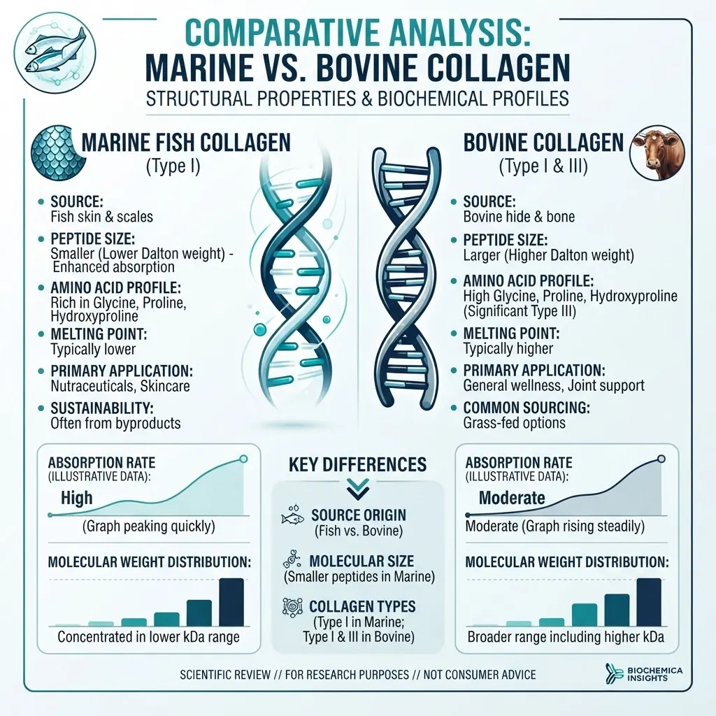
Research materials may be derived from bovine type I or III collagen, porcine collagen, marine type I collagen, recombinant collagen, or synthetic peptides. Each source has a different amino acid composition, molecular-weight distribution, and trace-impurity profile. Source selection can therefore affect assay interpretation and must be recorded with batch-specific documentation.
How do collagen peptide fragments interact with cells?
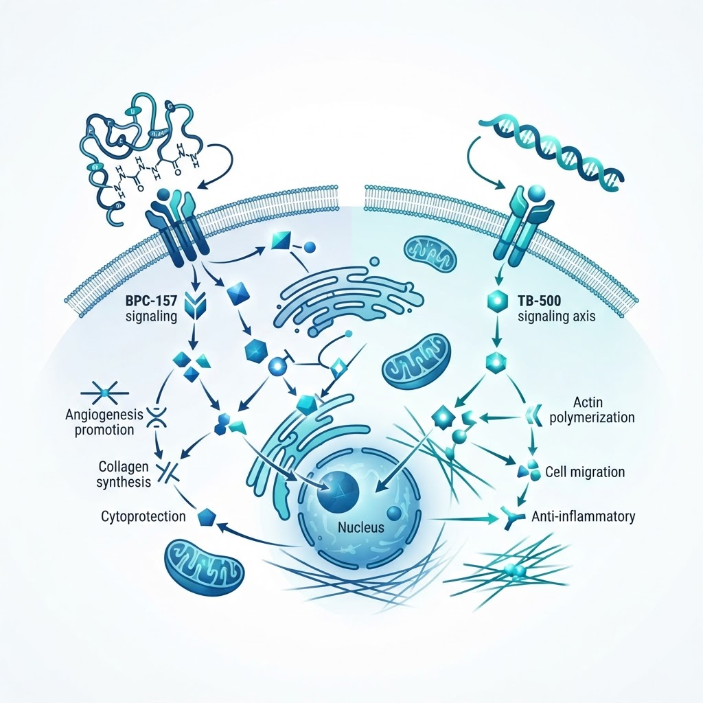
Collagen fragments interact with cells through receptor binding, extracellular-matrix association, and changes in the local chemical environment.
Integrin receptor binding
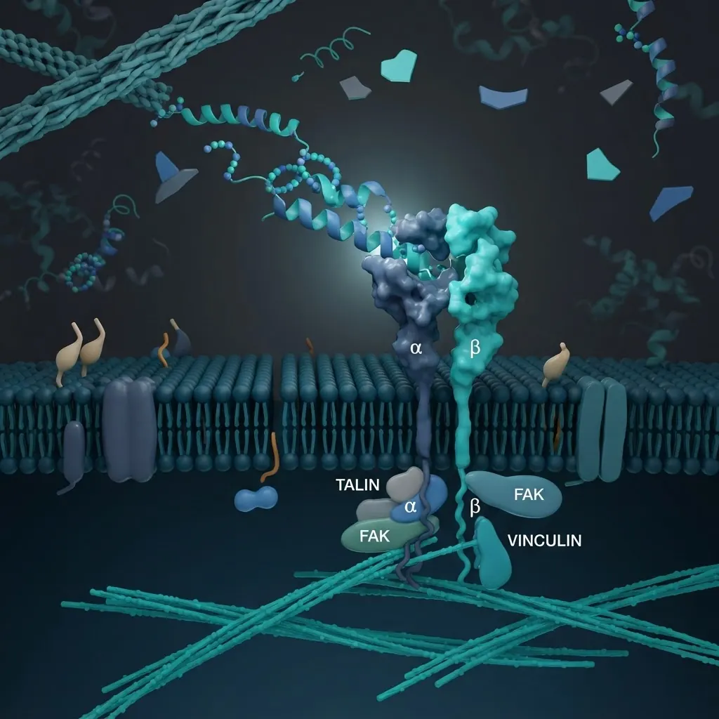
Specific collagen sequences bind integrin receptors on the cell surface. The GFOGER motif, found in type I collagen, is a well-established ligand for integrins including alpha1beta1 and alpha2beta1. Binding can recruit focal adhesion kinase and Src-family signalling, producing measurable changes in adhesion, spreading, migration, or matrix-gene expression.
| Integrin | Ligand motif | Common research readout |
|---|---|---|
| alpha1beta1 | GFOGER | Adhesion and matrix synthesis |
| alpha2beta1 | GFOGER | Adhesion and migration |
| alpha10beta1 | GFOGER | Chondrocyte adhesion |
| alpha11beta1 | Collagen motifs | Fibroblast adhesion |
Receptor specificity should be tested rather than inferred. Useful controls include scrambled sequences, receptor-specific inhibitors, uncoated surfaces, receptor knockdown, and comparison with full-length collagen.
Matrix interactions
Collagen fragments can also associate with fibronectin, laminin, proteoglycans, and other extracellular-matrix components. These interactions may alter cell attachment or the availability of other ligands. In complex mixtures, it is important to distinguish direct receptor binding from changes caused by adsorption, aggregation, or altered substrate presentation.
What collagen fragment classes are studied?
Type I fragments from skin, tendon, and bone are frequently studied in fibroblast adhesion, migration, and matrix-synthesis models. Type II fragments from cartilage are used in chondrocyte and cartilage-matrix research. Type III fragments are relevant to soft connective tissue models, while type IV fragments from basement membrane are used in endothelial adhesion and angiogenesis studies.
Hydrolysed collagen mixtures contain fragments across a broad molecular-weight range. Their aggregate effects can be useful experimentally, but the mixture must be characterised by chromatography and mass spectrometry before biological comparisons are made. A nominal label such as “collagen peptides” is not sufficient to establish sequence identity or distribution.
How are in-vitro connective tissue studies designed?
Cell model selection
Common models include dermal fibroblasts, chondrocytes, tenocytes, endothelial cells, and mesenchymal stem cells. Passage number, donor variability, serum lot, cell density, and differentiation state should be recorded. Chondrocytes can dedifferentiate in monolayer culture, while tenocytes may require mechanical stimulation for physiologically relevant interpretation.
Culture substrate
Fragments may be studied in solution or immobilised on a culture surface. For coating studies, researchers should define buffer composition, incubation temperature, coating time, surface chemistry, blocking conditions, and wash procedure. Uneven adsorption can produce apparent activity differences unrelated to sequence biology.
Timepoints and readouts
Adhesion is commonly measured over minutes to hours, migration over several hours to one day, matrix synthesis over one to three days, and differentiation over longer culture periods. Suitable readouts include adhesion staining, scratch or transwell migration, hydroxyproline quantification, qPCR for COL1A1 and COL3A1, MMP assays, and phospho-protein analysis. Multiple orthogonal readouts are preferable to a single endpoint.
What analytical methods verify collagen fragment identity?
Verification should cover identity, purity, concentration, composition, and molecular-weight distribution.
Amino acid analysis
Amino acid analysis can confirm characteristic glycine, proline, and hydroxyproline ratios. It is useful for assessing source consistency but does not identify every individual sequence in a hydrolysed mixture.
HPLC and mass spectrometry
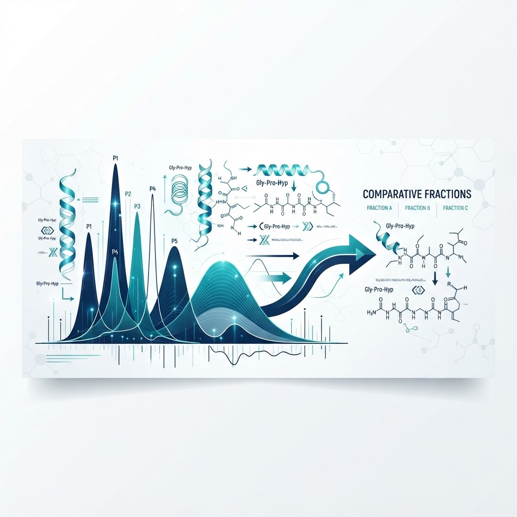
Reverse-phase HPLC separates fragments by hydrophobicity and reports purity as peak area under defined conditions. Mass spectrometry confirms molecular weight and, with tandem analysis, can provide sequence information. SEC can be used to determine molecular-weight distribution in complex hydrolysates.
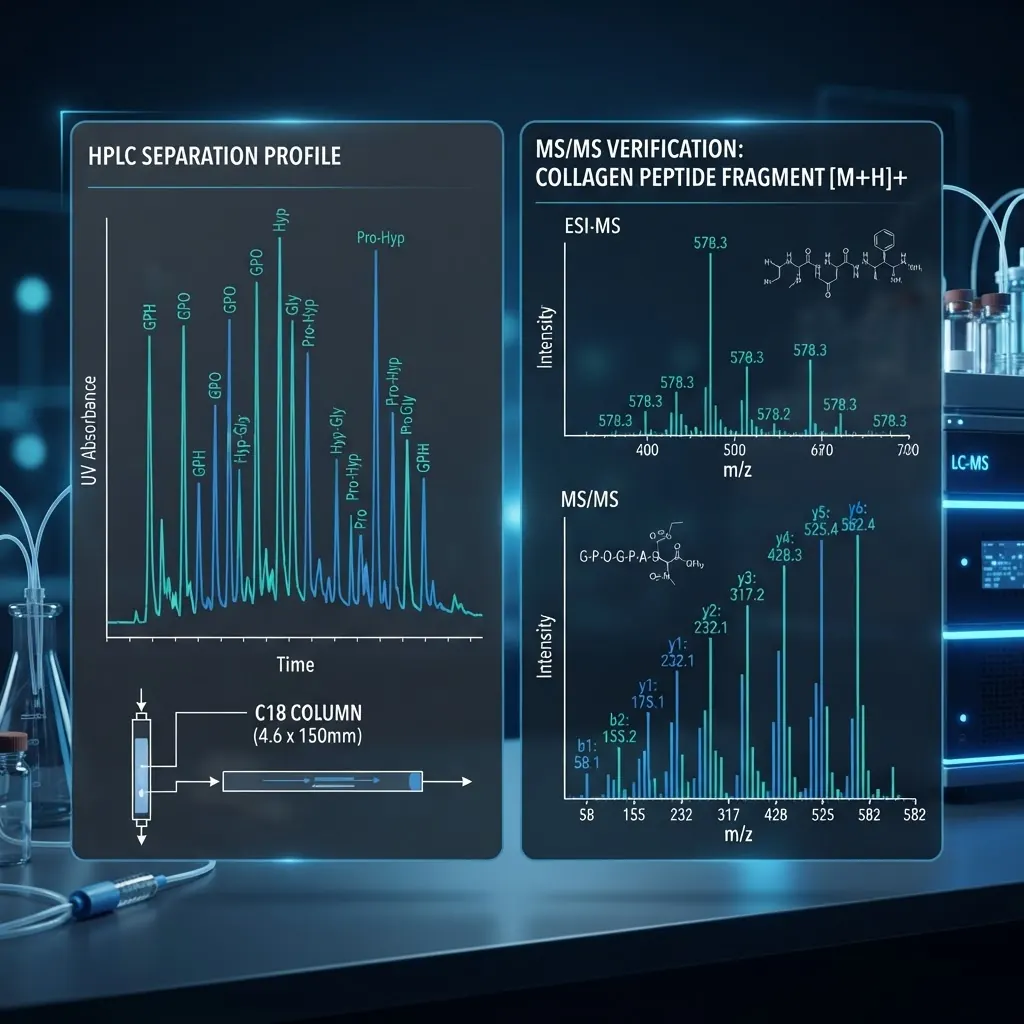
Hydroxyproline quantification provides an additional collagen-associated measure, but it should not replace chromatographic and mass-spectrometric characterisation. Batch-specific certificates, raw chromatograms, and method details improve reproducibility.
What stability variables should be controlled?
Fragment stability depends on sequence, concentration, buffer, temperature, protease activity, adsorption, oxidation, and aggregation. A stability study can incubate the material in the intended culture medium at 37°C, sample at defined intervals, and analyse the samples by HPLC or SEC. Low-binding plates and aliquoting can reduce adsorption and freeze-thaw artefacts.
Lyophilised material is generally stored sealed at low temperature. Reconstituted solutions should be aliquoted, clearly labelled, and evaluated for precipitation or concentration drift. Storage conditions must be validated for the specific sequence or mixture rather than copied from an unrelated material.
What are common research pitfalls?
Common sources of variability include uncharacterised source material, inconsistent coating, passage drift, serum-lot effects, unverified concentration, single-endpoint interpretation, missing scrambled controls, and failure to distinguish a defined synthetic sequence from a broad hydrolysate. Documentation should include batch identifiers, analytical methods, raw traces, cell passage, media composition, and surface preparation.
Applications in connective tissue research
Collagen fragments are used to study matrix remodelling, cell adhesion, migration, differentiation, and wound-model behaviour in vitro. They can help define how sequence, receptor engagement, matrix presentation, and analytical purity influence cellular measurements. Interpretation should remain limited to the model and readouts used; in-vitro activity does not establish clinical or animal outcomes.
For related methods, see [Peptide Signaling in Dermal Cell Research](/blog/peptide-signaling-dermal-cell-research), [Hydrolysed Collagen Peptides](/blog/hydrolysed-collagen-peptides-structure), and [HPLC Chromatography Guide](/blog/hplc-chromatography-guide).
Frequently Asked Questions
What are common collagen fragments used in research?
Gly-Pro-Hyp, Pro-Hyp, and the GFOGER motif are frequently studied sequence-defined examples. The appropriate fragment depends on the receptor, cell model, and assay.
How is fragment identity verified?
Use complementary amino acid analysis, HPLC, mass spectrometry, and, for mixtures, molecular-weight profiling by SEC or related methods.
What is the difference between hydrolysed collagen and a defined collagen peptide?
Hydrolysed collagen is a mixture with a distribution of fragment sizes. A defined collagen peptide has a specified sequence and molecular weight. They should not be treated as interchangeable test materials.
Which cells are used in connective tissue models?
Fibroblasts, chondrocytes, tenocytes, endothelial cells, and mesenchymal stem cells are commonly used, with selection driven by the tissue and endpoint.
How can batch variability be reduced?
Use one characterised batch where possible, retain raw analytical data, and verify equivalence before combining results from different batches.
Conclusion
Collagen peptide fragments are useful research tools for studying extracellular-matrix signalling and connective tissue cell behaviour. Reproducible work depends on defined material identity, suitable controls, validated analytical methods, and careful documentation of cell and culture variables. All compounds referenced are intended for in-vitro laboratory research only and are not for human or animal use.
References
- Shoulders MD, Raines RT. (2009). Collagen structure and stability. Annual Review of Biochemistry, 78, 929–958.
- Knight CG, Morton LF, Peachey AR, et al. (2000). Collagen-binding A-domains of integrins alpha1beta1 and alpha2beta1 recognize GFOGER. Journal of Biological Chemistry, 275(1), 35–40.
- Ricard-Blum S. (2011). The collagen family. Cold Spring Harbor Perspectives in Biology, 3(1), a004978.
- Heino J. (2007). The collagen receptor integrins have distinct ligand recognition and signalling functions. Matrix Biology, 26(7), 523–532.
Frequently asked questions
What are common collagen fragments used in research?
Gly-Pro-Hyp, Pro-Hyp, and the GFOGER motif are frequently studied sequence-defined examples. The appropriate fragment depends on the receptor, cell model, and assay.
How is fragment identity verified?
Use complementary amino acid analysis, HPLC, mass spectrometry, and, for mixtures, molecular-weight profiling by SEC or related methods.
What is the difference between hydrolysed collagen and a defined collagen peptide?
Hydrolysed collagen is a mixture with a distribution of fragment sizes. A defined collagen peptide has a specified sequence and molecular weight.
Which cells are used in connective tissue models?
Fibroblasts, chondrocytes, tenocytes, endothelial cells, and mesenchymal stem cells are commonly used.
How can batch variability be reduced?
Use one characterised batch where possible, retain raw analytical data, and verify equivalence before combining results from different batches.
Frequently Asked Questions
What are common collagen fragments used in research?
Gly-Pro-Hyp, Pro-Hyp, and the GFOGER motif are frequently studied sequence-defined examples. The appropriate fragment depends on the receptor, cell model, and assay.
How is fragment identity verified?
Use complementary amino acid analysis, HPLC, mass spectrometry, and, for mixtures, molecular-weight profiling by SEC or related methods.
What is the difference between hydrolysed collagen and a defined collagen peptide?
Hydrolysed collagen is a mixture with a distribution of fragment sizes. A defined collagen peptide has a specified sequence and molecular weight.
Which cells are used in connective tissue models?
Fibroblasts, chondrocytes, tenocytes, endothelial cells, and mesenchymal stem cells are commonly used.
How can batch variability be reduced?
Use one characterised batch where possible, retain raw analytical data, and verify equivalence before combining results from different batches.
Dr. James Whitfield
Our research team combines expertise in biochemistry, skincare science, and sports medicine to bring you evidence-based peptide information.
Ready to Explore Peptides?
Discover our selection of research-grade peptides and start your optimisation journey today.
Shop Peptides