# Marine vs. Bovine Collagen: Source Comparison for Laboratory Studies
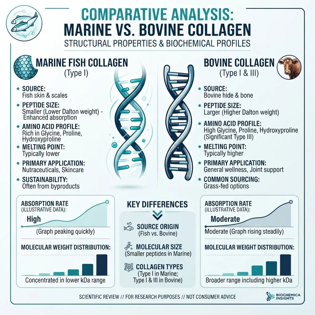
Collagen used in laboratory research is derived from multiple biological sources, most commonly marine fish tissues and bovine tissue. The source affects amino acid composition, molecular weight distribution, thermal stability, extraction profile, and performance in in-vitro models. These variables should be treated as experimental parameters rather than interchangeable product attributes.
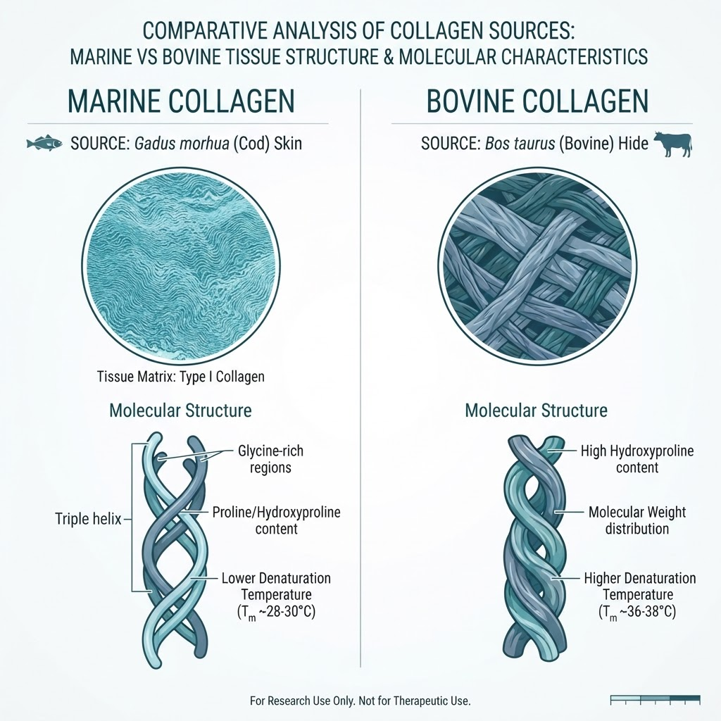
This technical comparison reviews source material, extraction methods, composition, analytical verification, and practical considerations for laboratory studies. All materials referenced are intended for in-vitro laboratory research only.
What are the primary sources of marine and bovine collagen?
Marine collagen is commonly extracted from fish skin, scales, and bone. Atlantic cod, salmon, and tilapia are frequent research sources. Fish skin generally provides type I collagen, while bone and cartilage can introduce additional collagen types and non-collagenous proteins. Jellyfish and sponge-derived materials are also studied, but their composition differs substantially from fish-derived collagen and should not be grouped together without verification.
Bovine collagen is commonly extracted from hide, tendon, bone, or cartilage. Bovine skin and tendon are predominantly type I collagen sources, while cartilage is associated with type II collagen. Source tissue, species, geographic origin, and extraction conditions all influence the final material. Bovine materials also require documented origin and transmissible spongiform encephalopathy controls appropriate to the intended research setting.
Acid-soluble and pepsin-soluble extraction are used for both source classes. Acid-soluble extraction can preserve more of the native triple-helical structure, while pepsin treatment can increase yield by removing terminal regions. Alkaline and targeted enzymatic processes may produce different fragment profiles. The extraction method should therefore appear in the sample record and certificate of analysis.
For a related structural overview, see [Collagen Peptide Fragments in Connective Tissue Research](/blog/collagen-peptide-fragments-connective-tissue).
How does amino acid composition differ between sources?
Both marine and bovine collagen are rich in glycine, proline, and hydroxyproline. Representative values vary by species and tissue, but marine collagen often shows approximately 6–10% hydroxyproline, while bovine collagen commonly falls around 9–13%. Glycine is typically close to one third of total residues in both materials. Proline, alanine, glutamic acid, and arginine also overlap considerably between sources.
Hydroxyproline is especially important because it contributes to triple-helix stability through hydrogen-bonding networks. A lower hydroxyproline content is one reason cold-water fish collagen often exhibits a lower denaturation temperature than bovine collagen. The difference is not universal: warm-water marine species, tissue selection, crosslinking, and processing can shift the observed values.
Amino acid analysis is therefore useful for source comparison, but it should not be used alone to identify species or tissue. LC-MS/MS peptide mapping, species-specific immunoassays, and orthogonal protein characterization provide stronger evidence when source identity matters.
How does molecular weight distribution compare?
Native type I collagen is approximately 300 kDa as a triple helix composed of three chains near 100 kDa each. Hydrolysis produces a distribution of shorter peptides rather than a single molecular species. The distribution depends on enzyme selection, reaction time, temperature, pH, and post-extraction filtration.
Research batches may show marine fragments broadly distributed from roughly 200 Da to 10 kDa, with many preparations showing a principal population around 1–3 kDa. Bovine hydrolysates may extend from approximately 300 Da to 15 kDa, with a principal population around 3–5 kDa. These values are indicative rather than specification limits; the batch-specific analytical trace should take precedence.
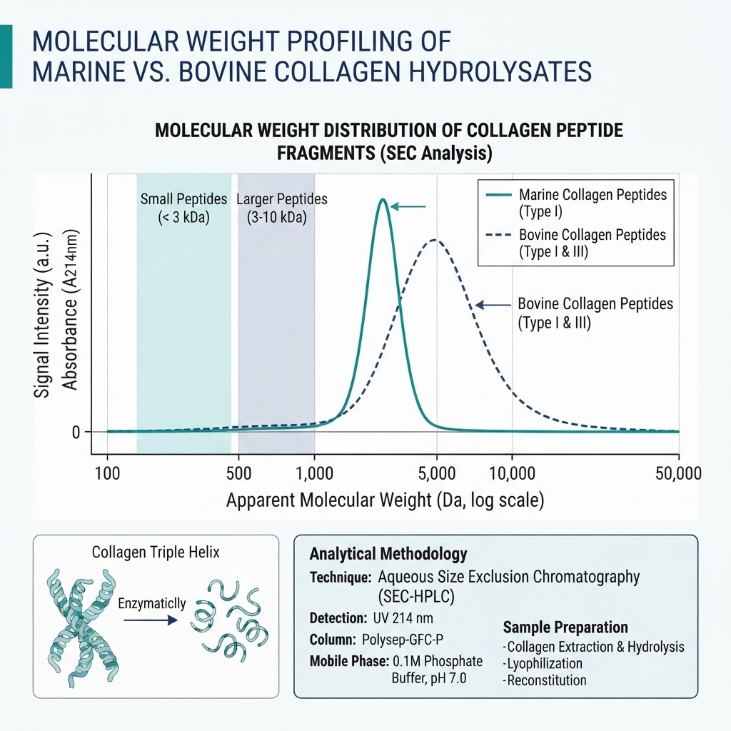
Lower-molecular-weight fragments generally diffuse more rapidly through membranes and may behave differently in cell-based assays. Larger fragments can retain more sequence context and may show different adsorption, aggregation, or receptor-interaction behaviour. Molecular weight should be reported alongside concentration, solvent, pH, and assay temperature because these variables can change the measured profile.
See [Hydrolysed Collagen Peptides: Molecular Structure and Research Applications](/blog/hydrolysed-collagen-peptides-structure) for additional characterization context.
What are the thermal stability differences?
Thermal stability is a critical experimental variable. The denaturation temperature, or Td, is the point at which the triple helix undergoes a helix-to-coil transition under defined conditions. Cold-water fish collagen may show Td values around 15–25°C, warm-water marine sources around 25–30°C, and bovine collagen commonly around 35–40°C. Exact values depend on species, tissue, pH, ionic strength, and measurement method.
A marine preparation that denatures near room temperature may not preserve native structure during a 37°C cell-culture experiment. This does not make it unsuitable for research, but it means that the experimental question should distinguish native-structure effects from effects produced by denatured collagen or peptide fragments. Storage, thawing, and reconstitution should likewise be controlled and documented.
Circular dichroism can monitor loss of helical signal as temperature increases. Differential scanning calorimetry measures the heat absorbed during denaturation. Viscosity changes can provide a complementary indication of the helix-to-coil transition. Researchers should verify the thermal profile of the actual batch rather than relying only on a source label.
What analytical methods verify collagen source and quality?
A defensible source comparison uses several orthogonal methods. Amino acid analysis quantifies residue composition and can identify broad differences in hydroxyproline content. A hydroxyproline assay estimates total collagen in a mixture, but it does not establish species identity. SDS-PAGE helps assess intact alpha, beta, and gamma chains and larger fragments.
Reverse-phase HPLC separates peptide fragments according to hydrophobicity and can reveal differences between batches. Size-exclusion chromatography provides a more direct view of molecular weight distribution. LC-MS/MS identifies peptide sequences and can support species assignment, while MALDI-TOF provides rapid mass profiling for suitable fragment mixtures. Circular dichroism is useful where triple-helix content is an experimental variable.
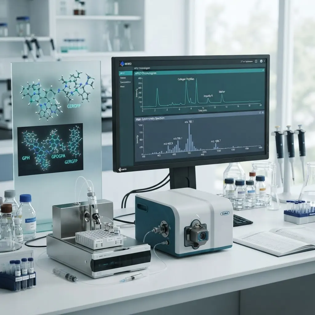
For method selection, see the [HPLC Chromatography Guide](/blog/hplc-chromatography-guide) and [Peptide Purity Testing Standards](/blog/peptide-purity-testing-standards). A complete sample record should include source species, tissue, extraction chemistry, hydrolysis conditions, purity, molecular weight trace, amino acid profile, endotoxin result, and storage history.
What are the regulatory and safety considerations for research materials?
Bovine materials should be accompanied by country-of-origin information, BSE or TSE documentation where applicable, and species verification. Marine materials should include species documentation, allergen information for fish-derived sources, and heavy-metal testing appropriate to the source environment. These records support traceability and help researchers interpret unexpected assay variation.
Endotoxin testing is particularly important for cell-culture work. Limulus amoebocyte lysate assays are commonly used, although the selected method should be compatible with the sample matrix. A commonly cited research threshold is below 1 EU per microgram, but the acceptable limit depends on the cell system and study design. Researchers should define acceptance criteria before beginning the experiment.
What are the practical considerations for in-vitro research?
Marine collagen is often more soluble at lower temperatures because of its lower hydroxyproline content. Bovine collagen may require acidic conditions or controlled warming for dissolution. Neither source should be assumed to have the same coating efficiency, adsorption behaviour, or fibril formation kinetics. Coating concentration, surface chemistry, incubation time, and wash steps should be kept consistent when comparing sources.
Both sources can support cell-adhesion and matrix-model experiments, but cell responses are model-dependent. A mammalian cell line may interact differently with marine and bovine sequences, and a fish-derived model may produce a different result again. Include an uncoated control, record passage number and cell density, and avoid attributing a source effect to collagen without checking pH, osmolality, endotoxin, and residual solvent.
Store sealed research materials according to the supplier specification, commonly at -20°C or -80°C. Limit freeze-thaw cycles and record deviations. Request a batch-specific certificate of analysis documenting the molecular weight distribution, composition, purity, and microbiological testing. Our [Quality Assurance page](/quality-assurance) lists batch documentation.
For broader handling considerations, see [Peptide Storage and Stability Best Practices](/blog/peptide-storage-stability-science-backed-best-practices).
How do you select the right source for a study?
Select marine collagen when the study concerns fish-derived materials, cold-water biology, lower thermal stability, or source-dependent peptide diffusion. Select bovine collagen when mammalian tissue comparability, higher thermal stability, or a well-characterized type I and type III background is important. Use both when the study is explicitly comparative or investigates species-dependent receptor interactions.
The source should be selected after defining the endpoint. If the endpoint is triple-helix retention, thermal analysis and structure-preserving extraction are central. If the endpoint is fragment response in a cell assay, molecular weight distribution, sequence identity, endotoxin, and solvent controls may be more important. A source decision is therefore a study-design decision, not a ranking.
What are common pitfalls in collagen source research?
Common reproducibility problems include unverified species or tissue, assuming that two hydrolysates have the same molecular weight profile, ignoring denaturation temperature, mixing batches, and failing to test endotoxin or heavy metals. Other issues include incomplete extraction records, mismatched solvent conditions, cell-line differences, and temperature excursions during storage.
Avoid these issues by defining the material specification in advance, testing every new batch against the specification, and reporting the relevant analytical traces with the experimental results. Do not pool data across marine and bovine sources unless the study is designed to test source effects.
Conclusion
Marine and bovine collagen differ in amino acid composition, molecular weight distribution, extraction profile, and thermal stability. Marine materials often show lower hydroxyproline content and lower denaturation temperatures, while bovine materials commonly provide greater thermal stability. Neither source is universally preferable: the appropriate choice depends on tissue origin, model system, endpoint, and the analytical controls used.
All materials referenced in this article are intended for in-vitro laboratory research only. They are not medicines, supplements, or consumer products, and are not for human or animal use.
Frequently Asked Questions
What is the main difference between marine and bovine collagen?
Hydroxyproline content and thermal stability are common points of difference, but the result varies by species, tissue, and processing. Source identity should be confirmed with orthogonal analytical methods.
Why can marine collagen have lower thermal stability?
Many cold-water fish collagens contain less hydroxyproline, which can reduce stabilization of the triple helix. The measured Td is batch- and method-dependent.
Can the sources be used interchangeably in cell culture?
Not without verification. Molecular weight, sequence, thermal behaviour, endotoxin, and coating properties can differ and may affect the cell response.
How is source identity verified?
Use amino acid analysis, peptide mapping by mass spectrometry, SDS-PAGE, and species-specific assays as appropriate for the study.
What is the typical molecular weight?
Native collagen is near 300 kDa, while hydrolysed preparations contain broad fragment distributions. SEC or SDS-PAGE should be used to characterize the actual batch.
How should the samples be stored?
Follow the batch specification, commonly using sealed containers at -20°C or -80°C and minimizing freeze-thaw cycles.
Which source is appropriate for mammalian models?
Bovine collagen may provide closer tissue comparability for some mammalian models, but the choice should be justified by the endpoint and verified experimentally.
When should both sources be compared?
Compare both when the study asks a source-dependent question, such as thermal stability, sequence-specific interaction, or species-related cell response.
Frequently asked questions
What is the main difference between marine and bovine collagen?
Hydroxyproline content and thermal stability are common points of difference, but the result varies by species, tissue, and processing.
Why can marine collagen have lower thermal stability?
Many cold-water fish collagens contain less hydroxyproline, which can reduce stabilization of the triple helix.
Can the sources be used interchangeably in cell culture?
Not without verification because molecular weight, sequence, thermal behaviour, endotoxin, and coating properties can differ.
How is source identity verified?
Amino acid analysis, mass spectrometry, SDS-PAGE, and species-specific assays can be used as appropriate.
What is the typical molecular weight?
Native collagen is near 300 kDa, while hydrolysed preparations contain broad fragment distributions.
How should the samples be stored?
Follow the batch specification, commonly using sealed containers at -20°C or -80°C and minimizing freeze-thaw cycles.
Which source is appropriate for mammalian models?
Bovine collagen may provide closer tissue comparability for some mammalian models, but the choice should be justified by the endpoint.
When should both sources be compared?
Compare both when the study asks a source-dependent question, such as thermal stability or species-related cell response.
Frequently Asked Questions
What is the main difference between marine and bovine collagen?
Hydroxyproline content and thermal stability are common points of difference, but the result varies by species, tissue, and processing.
Why can marine collagen have lower thermal stability?
Many cold-water fish collagens contain less hydroxyproline, which can reduce stabilization of the triple helix.
Can the sources be used interchangeably in cell culture?
Not without verification because molecular weight, sequence, thermal behaviour, endotoxin, and coating properties can differ.
How is source identity verified?
Amino acid analysis, mass spectrometry, SDS-PAGE, and species-specific assays can be used as appropriate.
What is the typical molecular weight?
Native collagen is near 300 kDa, while hydrolysed preparations contain broad fragment distributions.
How should the samples be stored?
Follow the batch specification, commonly using sealed containers at -20°C or -80°C and minimizing freeze-thaw cycles.
Which source is appropriate for mammalian models?
Bovine collagen may provide closer tissue comparability for some mammalian models, but the choice should be justified by the endpoint.
When should both sources be compared?
Compare both when the study asks a source-dependent question, such as thermal stability or species-related cell response.
Dr. Sarah Chen
Our research team combines expertise in biochemistry, skincare science, and sports medicine to bring you evidence-based peptide information.
Ready to Explore Peptides?
Discover our selection of research-grade peptides and start your optimisation journey today.
Shop Peptides